Digging Lungfish – The Biological Miracle Dormant in Mud


A lungfish underwater

A Lungfish (Protopterus annectens) at Paris Aquarium.

Now imagine this – it was just an ordinary day in West Africa. Under the scorching summer sun, you were building yourself a hut with mud bricks made out of the dried ground. Life was peaceful until one night, when there was an unusually heavy rain. You, soaked in rainwater, sadly watched the mud being washed off your home, but were soon horrified – snake-like creatures were coming out of the bricks and dragging themselves down the walls! Now, don’t panic, because you just witnessed one of the most amazing animals on Earth – the lungfish, who have been asleep in your mud walls for months.

The African lungfish, genus Protopterus, are “living fossils” that have survived on the Earth for nearly 400 million years, through drastic environmental changes such as the drying of the Sahara. One of the main adaptations that allows lungfish to be the ultimate survivor is aestivation – dormancy in torrid seasons (as opposed to hibernation in winter), for up to an impressive 4 years.

Your encounter in Africa is not entirely hypothetical. And, believe it or not – people in Africa have even been digging lungfish out of dry mud for food.

A lungfish with half of its body buried in moist mud

West African lungfish (Protopterus annectens) buried in mud of dried river bed, Togo.

So, how can an aquatic fish live in dry mud without any food or water, even sleeping in your wall for months without disturbance? To answer this question, curious scientists have been observing African lungfish since the 20th century.

In 1986, scientists from the University of Pennsylvania School of Medicine recorded the behavior of aestivating lungfish. Aestivation happens in dry seasons, as the retreating water leaves lungfish on moist mud. Without warning, lungfish starts to burrow a hole into the mud, and periodically resurfaces to take big gulps of air. It repeats this process until it’s lodged inside the hole in an upright U shape, with its mouth placed next to a thin channel in the ground for breathing.

a U-shaped lungfish in its hole in the ground, with its mouth next to a thin channel connected to the ground

A computer-generated model of a lungfish in its burrow.

By this time, lungfish has excreted enough tenacious mucus from its skin, which hardens to form a bed – a cocoon that envelops the lungfish’s entire body. Perhaps more unfortunate than starving, lungfish doesn’t get to turn in its sleep. It would remain this posture until enough water finally triggers its awakening.

With dissection and microscopy, scientists from the University of Genoa looked inside the amazing fish. To adapt to a new life underground, lungfish changes both the structure and function of its organs.

Most obviously, it transitions from water-breathing to air-breathing on land. True to its name, lungfish has both good old gills and lungs that resemble the lung types of primitive amphibians.

The dissected inside of a lungfish, showing its left lung, notochord, and liver.

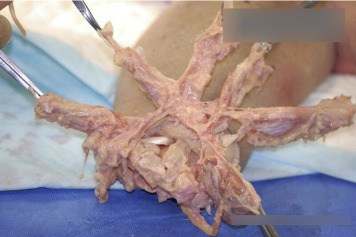
Lateral view of lungs of a Protopterus dolloi, from lab dissection at U. of Cincinnati.

In water, lungfish’s gills have separated filaments with large surface area, increasing the gas exchange between blood and water. When on land, however, the aestivating fish excretes mucus that makes gill filaments stuck together, therefore non-functional.

On the contrary, the fish’s lungs that are thin bloodless threads in water are now swollen with alveoli – tiny “balloons” that transport gas into and out of the bloodstream. In this way, lungfish can breathe like a turtle.

Heart and circulation of the lungfish - when estivating, its blood predominantly flows through the lungs; when underwater, its blood predominantly flows through the gills instead.

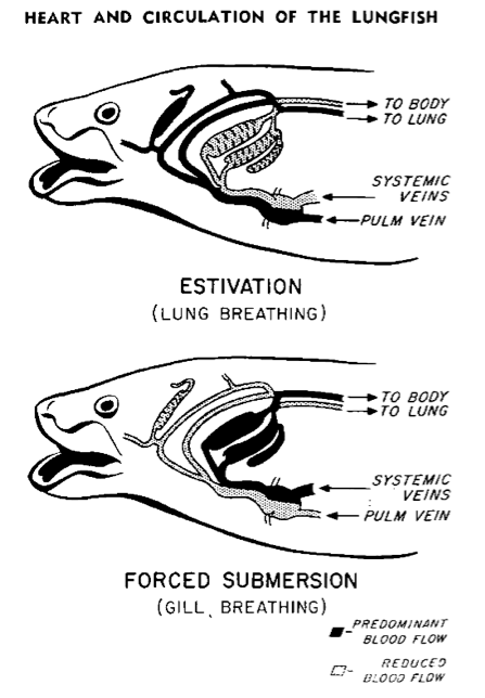
Illustration of lungfish’s blood flow accommodating different breathing modes.

In recent years, development of genetic tools has revealed more subtle changes inside the lungfish’s body. Through compiling genes and examining the expression levels of different DNAs, scientists from the National University of Singapore have found that lungfish adjusts its metabolism to resist stress and save energy.

For instance, apart from breathing, the aestivating fish shuts down another function of gills – excreting toxic waste as ammonia into the water. When on land, the lack of water makes this way of excretion impossible. To save energy, lungfish decreases the production of gill-proteins responsible for ammonia transport. It instead uses liver to convert ammonia into the less toxic urea.

The miracle of lungfish biology, however, does not stop with these clever mechanisms for dormancy. I can easily name a few more reasons for why lungfish is the coolest animal the Earth has ever seen.

(1) Lungfish has lots, lots of genes. The marbled lungfish from Africa (Protopterus aethiopicus) has the second largest genome – the total genetic material of an organism – found by scientists so far, amongst all species in the world. The only larger one belongs to a Japanese flower named Paris japonica.

(2) We are related. The similarity in physiology and gene sequences assigns lungfish as the closest living relative among fish of land vertebrates.

(3) Aestivation matters. From aestivation, hibernation, to tardigrades’ ability to shrivel up into a ball and survive in space, these common yet diverse forms of dormancy are called suspended animation. Scientists are now hypothesizing that there is a common genetic base for this ancient adaptation, which is likely to be also present in humans.

One essential question left unanswered, however, is the exact factors that trigger organisms into and out of dormancy. By potentially harnessing this “switch” of suspended animation, we can make ourselves survive in far harsher environments.

For patients awaiting organ transplants, this technology may be a life-savior; for astronomers, this raises hope to look for dormant life forms in outer space, or even put astronauts into dormancy for space exploration. Aestivation, as one unique form of dormancy found in an organism of evolutionary significance, is definitely worth more studying.

An astronaut falls asleep inside a box filled with liquid. The lid closes slowly.

Astronauts entering “hypersleep” on the way to Saturn, from Nolan’s 2014 sci-fi epic Interstellar. Could this be true one day?

Before we can finally put cancer patients to a temporary sleep or send snoozing astronauts into deep space, let’s look at unexpected places for inspiration. Hidden underground, much more miracles of nature are there for us to see.

By: Z Zhang

References

Alekseev V.R., Sychev V.N., Novikova N.D. (2007). Studying the Phenomenon of Dormancy: Why it is Important for Space Exploration. Diapause in Aquatic Invertebrates Theory and Human Use. Monographiae Biologicae, (84), 207-214. Springer, Dordrecht. Retrieved from https://link.springer.com/chapter/10.1007%2F978-1-4020-5680-2_14

Chng, Y. R., Ong, J. L. Y., Ching, B., Chen, X. L., Hiong, K. C., Wong, W. P., . . . Ip, Y. K. (2017). Molecular characterization of three rhesus glycoproteins from the gills of the african lungfish, protopterus annectens, and effects of aestivation on their mRNA expression levels and protein abundance. PLoS One, 12(10), e0185814. Retrieved from http://dx.doi.org/10.1371/journal.pone.0185814

Fishman, A. P., Pack, A. I., Delaney, R. G., & Galante, R. J. (1986). Estivation in Protopterus.  J. Morphol., 190(S1), 237–248. Retrieved from https://doi.org/10.1002/jmor.1051900416

Hiong, K. C., Ip, Y. K., Wong, W. P., & Chew, S. F. (2015). Differential gene expression in the liver of the african lungfish, protopterus annectens, after 6 months of aestivation in air or 1 day of arousal from 6 months of aestivation. PLoS One, 10(3), e0121224. Retrieved from http://dx.doi.org/10.1371/journal.pone.0121224

Sturla, M., Paola, P., Carlo, G., Angela, M. M., & Maria, U. B. (2002). Effects of induced aestivation in Protopterus annectens: A histomorphological study. J. Exp. Zool., 292(1), 26–31. Retrieved from https://doi.org/10.1002/jez.1139

Image Credits

Image 1: Mathae, Protopterus annectens at Paris Aquarium,  https://commons.wikimedia.org/wiki/File:G%C5%91tehal-2.jpg

Image 2: Heuclin, D., West African lungfish (Protopterus annectens) buried in mud of dried river bed, Togo., //www.naturepl.com/stock-photo-nature-image01597946.html

Image 3: Unknown author, //lh3.googleusercontent.com/-FRv0ocgqMpU/WJw2hiUhcXI/AAAAAAABRtY/xxIvkiQzf5k/lungfish-52.jpg?imgmax=1600

Image 4: Mokele, Lungs of Protopterus dolloi, https://commons.wikimedia.org/wiki/File:Lungs_of_Protopterus_dolloi.JPG

Image 5: Szidon, Jan P., Figure 16 from Heart and Circulation of the African Lungfish, https://www.ahajournals.org/doi/abs/10.1161/01.res.25.1.23

Image 6: Interstellar (2014), Romilly entering hypersleep on the way to Saturn, https://www.imdb.com/title/tt0816692/

Therapeutic Advantages of Embryonic Stem Cells

Embryonic Stem cells, Cellular Therapy

Research on human stem cells has achieved a lot since the cloning of Dolly the lamb and the identification of the human genome sequence. Stem cells can now be personalized and developed into specific cells for specific needs, and have developed an immense potential to help patients suffering from various conditions. This progress has ultimately created suspicion and awareness of ethical issues that can arise from this science.

The possibility of reproductive human cloning and acquisition of embryos through unjust means have prompted countries including Germany, Austria, Italy and many others to ban or restrict research in the stem cell field. Although the ethical issues surrounding this research are appropriate, stem cells can be used in very beneficial ways.

One method of generating specialized and personalized stem cells, called somatic cell nuclear transfer, can be used for therapeutic treatment and for further research of genetic diseases. This cloning technique of somatic-cell nuclear transfer can be used to successfully generate embryonic stem cells. Scientist Shoukrat Mitalipov from the Oregon National Primate Research Center, has generated positive results through research and studies of producing monkey and human embryos.

Mitalipov’s work has proven to create embryonic stem cells when caffeine is added to the somatic-cell nuclear transferred embryos. His method yields that 23% of somatic cell nuclear transferred embryos will form into blastocysts, which are embryos that have developed hundreds of cells. These blastocysts, can then be used to generate embryonic stem cells. And these embryonic stem cells can then become personalized or specialized according to needs of patients.

This development would be very beneficial therapeutically, but still raises concerns over reproductive cloning. But contrary to many beliefs that embryonic stem cells could be cultivated and used to clone humans, the current state of this science holds that these cells have little to no chance of further development. Mitalipov also performed studies on monkeys in order to resolve this belief. His study found that

“despite more than 100 tries, he and his colleagues have not managed to get any SCNT-derived monkey embryos to implant in a surrogate mother and trigger pregnancy. The cells that give rise to the placenta are underdeveloped in the cloned monkey embryos, he says, and the human SCNT embryos have similar abnormalities.”

These efforts provide evidence of how the current state of stem cells would not entertain reproductive cloning and if properly overseen this method of achieving embryonic stem cells remains appropriate.

This method as mentioned before, can be very useful for therapeutic treatment. A few areas it can prove very useful therapeutically include

“Restoring bone growth after bone injury. Restoring vision in retinal disease. Restoring nerve cell function in spinal cord injury. Restoring heart tissue after heart attack,”

and many genetic diseases such as Parkinson’s, Alzheimer’s, and cancers. One particular treatment embryonic stem cells can prove especially beneficial for is treatment for spinal cord injuries.

Skeletal Human Spine

According to reports by Vibhu Sahni and John A. Kessler published by the National Center for Biotechnology Information ,

“ES cells have been shown to have therapeutic benefits only when transplanted into experimental models of mild to moderately severe SCI (Spinal Cord Injury).”

This development would significantly help the countless people effected by spinal cord problems, although this treatment is not as effective for major spinal cord injuries. The number of Americans alone that suffer from spinal cord injury is substantial. Sahni and Kessler report that

“A 2004 study sponsored by the Christopher and Dana Reeve Foundation revealed that 1,275,000 people in the US have some form of SCI.”

These numbers reinforce the need for more research and efforts towards spinal cord injuries and this form of treatment, as it can replace current therapy and would require less drugs in the process.

A biotechnology company based in California, called Geron Corporation performed studies on embryonic stem cell treatment for spinal cord injuries in 2007. Their research studies not only showed that embryonic stem cells can treat this problem, but can also avoid attack from the immune system. According to Thomas B. Okarma, president of Geron, this treatment avoids recognition from the immune system. A different treatment such as a solid organ transplant would experience attacks in the form of lysis by the immune system, which is the destruction of cells through a process of breaking the cell membrane. These types of treatments require considerable immunosuppression drugs to allow the treatment to proceed, but with embryonic stem cells the immune system has a much less aggressive response.

This development is notable as the need for immunosuppression drugs, typically required for other spinal cord treatments in large doses, is not as significant for embryonic stem cell treatment. Lowered dosage of immunosuppression drugs for patients would benefit them greatly as it

“decreases the potential for untoward side effects that are common with those types of drugs” according to studies by Geron.

Side effects of these drugs include trembling, fever, infection, coughing, and many other symptoms. Thus, stem cell treatment for spinal cord injuries adequately provides therapy for this problem, while also reducing the risks that other forms of treatment pose through post-surgical therapy.

The ethical concerns revolving around stem cell research will remain valid. Yet in the current state of this science, there is nearly zero chance of human reproduction through stem cells, and along with proper regulation and oversight these issues would not be debated. Embryonic stem cells offer to help numerous patients and allow scientists to study and learn more about genetic diseases. It can provide therapeutic treatment for tissue, retina, and bone marrow restoration, and promote further studies of genetic diseases like Parkinson’s or Alzheimer’s. If this field is given serious support, and appropriate regulation and oversight, embryonic stem cells can help the countless suffering patients and make certain drugs obsolete.

By: S. Loyack


References

Bae, Sun Hwan. “Recent Achievements in Stem Cell Therapy for Pediatric Gastrointestinal Tract Disease.” Pediatric Gastroenterology, Hepatology & Nutrition, The Korean Society of Pediatric Gastroenterology, Hepatology and Nutrition, Mar. 2013, www.ncbi.nlm.nih.gov/pmc/articles/PMC3746046/

“EIExplorationInvestor: Geron Reports Studies Indicate that Human Embryonic Stem Cell-Based Therapeutic Treatment of Spinal Cord Injury Evades Attack by Human Immune System.” M2 Presswire, Normans Media Ltd, 11 June 2007, http://libproxy.lib.unc.edu/login?url=https://search.proquest.com/docview/444688057?accountid=14244

Gupta, P. N. “10 Things You Should Know About Immunosuppressants.” Organ Donation India, Organ Receiving and Giving Awareness Network, 15 Oct. 2019, http://www.organindia.org/ORGAN-DONATION/10-things-you-should-know-about-immunosuppressants/.

Lo, Bernard, and Lindsay Parham. “Ethical Issues in Stem Cell Research.” OUP Academic, Oxford University Press, 1 May 2009, http://academic.oup.com/edrv/article/30/3/204/2354990.

“NCI Dictionary of Cancer Terms.” NCI Dictionary of Cancer Terms, National Cancer Institute, www.cancer.gov/publications/dictionaries/cancer-terms/def/lysis.

Sahni, Vibhu, and John A Kessler. “Stem Cell Therapies for Spinal Cord Injury.” Nature Reviews. Neurology, U.S. National Library of Medicine, July 2010, http://www.ncbi.nlm.nih.gov/pmc/articles/PMC3755897/.

“Stem Cells.” MedlinePlus, U.S. National Library of Medicine, 20 Dec. 2019, http://medlineplus.gov/stemcells.html.

Vogel, Gretchen. “Human Stem Cells From Cloning, Finally.” Science, American Association for the Advancement of Science, 17 May 2013, http://science.sciencemag.org/content/340/6134/795.full


Image Credits

Image 1: Cancemi, Giovanni, “Embryonic Stem Cells, Cellular Therapy.” Adobe. https://stock.adobe.com/183494901?as_campaign=TinEye&as_content=tineye_match&epi1=183494901&tduid=7d4822674b887e5394f68f7c73d67b77&as_channel=affiliate&as_campclass=redirect&as_source=arvato

Image 2: “Skeletal Human Spine.” Adobe. https://stock.adobe.com/187133151?as_campaign=TinEye&as_content=tineye_match&epi1=187133151&tduid=7d4822674b887e5394f68f7c73d67b77&as_channel=affiliate&as_campclass=redirect&as_source=arvato

Inequalities on the UNOS Transplant Lists Have Gone On for Too Lung

By: R. Peterken

//rightasrain.uwmedicine.org/well/health/how-organ-donation-works

The Gift of Life by Ada Love

What would you do if a loved one’s only chance at living was getting a high-demand organ that’s in low supply? The lack of supply of organs versus the demand for them is one of the biggest current issues with the United Network for Organ Sharing (UNOS) waitlists (there are separate waitlists for children and adults).

There is much debate about how to ethically rank patients for receiving an organ, especially because different people have varying opinions on what factors are important.  According to the official UNOS website, factors like blood type and age contribute to where a patient is placed on their respective waitlist for all donated organs. However, there are also factors that are specific to the certain organ that is needed.  This piece will focus mainly on those patients who need liver transplants and what factors contribute to their ranking as well as the inequalities that they face.  Important factors specific to liver transplant patients on the UNOS lists include medical urgency and the patient’s Model for End Stage Liver Disease (MELD) score.  According to the journal Annals of Gastroenterology, patients who have a higher MELD score typically are on the wait lists for less time.    

While the UNOS transplant lists have benefits for many liver transplant patients, they also have major pitfalls. The official UNOS website explains that the lists currently are created by a computer that receives both donor and transplant data.  The computer takes the information and makes a ranked list of patients who could potentially get the organ based off general (ex. blood type) and organ specific factors (ex. medical urgency).  One paramount benefit of this system includes that a computer is able to rank the patients, meaning that healthcare and administrative personnel do not have to make the hard decision of how organs are rationed.

Another pro to the UNOS ranking system is that children and adults are each given their own specific list, meaning the organs can be given to the age group/patients that will have the best chance of a successful transplant and recovery.  On the other hand, a pitfall is the apparent inequalities that will later be discussed involving the system of ranking patients/ the UNOS lists.   Inequalities can be shown by investigating which patients get transplants and how this correlates to factors such as ethnicity, socioeconomic status, and age.

Inequalities can quickly become harmful and even deadly for many patients who need liver transplants if they are unable to obtain an organ before succumbing to their illness. A study from Gastroenterology, vol. 154, explored data on patients removed from the UNOS adult wait list in the U.S. from 2006-2016 due to receiving an organ transplant, death, etc. The findings showed that there were several inequalities stemming from influences such as insurance type.

This same journal stated that patients who do not have private insurance are more likely to die while still on the waiting list.  According to a section from a different journal, Gastroenterology and Hepatology, the National Healthcare Disparities Report said that not having insurance was a huge obstacle in getting healthcare for both Asian and African American populations.  Having private insurance makes it much easier to get evaluated for UNOS waitlist candidacy and can even get patients evaluated at multiple centers. While it is beneficial to have, is not always possible to obtain because of the high costs.

Another major inequality discussed in a study in the journal Gastroenterology, vol. 154 was the fact that Black and Asian patients were found to be less likely than whites to die on the waiting lists.  While this may at first seem like an inequality to whites, the official current numbers of each ethnicity on the UNOS waiting lists tell a different story.  The data shows that the number of white patients registered as candidates for a liver transplant is about the same as the number of patients from all other ethnicities combined.  This means that while more whites are dying, there are thousands more of them on the UNOS lists for liver transplants compared to other ethnicities.

One explanation for why there are so few minorities in the U.S. on the list is that liver transplants are extremely expensive, and many patients who are Asian, Hispanic, etc. may not have the means to be able to afford getting evaluated, medical bills or transfers to other centers when organs become available.  Inequalities such as the ones stemming from being a minority cause a large amount of skepticism for UNOS and its current system.

The skepticism previously mentioned means that many people are scared to donate organs, which breeds a cycle of organ shortage.  In fact, in a study from the early 2000’s discussed in the Journal of General Internal Medicine, it was stated that many populations (especially African Americans) were either unwilling to donate or did not understand how great the shortage for livers was.  Many also did not trust the healthcare system in general because of the inequalities present.

According to journals such as the Revista de Bioteca y Derecho, one way to lessen the inequalities on the lists would be to make the UNOS lists public.  This journal explains that doing so would lead to less skepticism in the general public.  This is because if the public was able to see where patients were on the lists, they would be more likely to believe that the lists would not be unfairly changed to help certain races, celebrities, etc.  This would also make it possible to see some of the inequalities that minorities face and could lead to policy changes that help solve these problems.  A build-up of trust and lessening of inequalities would increase the amount of organs being donated, essentially lowering the gap between the supply and demand.

This research shows that while the current UNOS system used does have its pros, it is defined by many to be a broken system.  Without change, there will continue to be a high volume of patients who die without the organ transplant that they need. Making the list transparent is just one way that would likely increase the public’s trust in UNOS, and make many more willing to donate.  This would progressively lessen the cycle of inequality that stems from a lack of organs. Discussions surrounding the issues on the UNOS organ transplant lists and possible solutions need to become more prominent for patients in the United States to have a happier, healthier future.

References:

“How We Match Organs.” UNOS, unos.org/transplant/how-we-match-organs/. 10 February 2020.

Kemmer, Nyingi. “Ethnic disparities in Liver Transplantation.” Gastroenterology & Hepatology vol. 7, (2011). https://www.ncbi.nlm.nih.gov/pmc/articles/PMC3127035/.

“Organ Procurement and Transplantation Network.” OPTN, OPTN, optn.transplant.hrsa.gov/data/view-data-reports/national-data/#. 11 February 2020.

Shen, Nicole T., et al. “Sa1508 – National and Regional Ten Year Assessment of United Network for Organ Sharing (UNOS) Liver Transplant Data Wait List Removals: Improvement over Time but Inequalities Persist.” Gastroenterology, vol. 154, no. 6, 2018, p. S1137.,https://www.sciencedirect.com/science/article/pii/S0016508518337752

Siminoff, Laura A et al. “Racial Disparities in Preferences and Perceptions Regarding Organ Donation.” Journal of General Internal Medicine vol. 21,9 (2006): 995-1000. https://www.ncbi.nlm.nih.gov/pmc/articles/PMC1831604/

Trieu, Judy A et al., “Factors associated with waiting time on the liver transplant list: an analysis of the United Network for Organ Sharing (UNOS) database.” Annals of gastroenterology vol. 31, 155, 1 (2018): 84-89.http://www.annalsgastro.gr/files/journals/1/earlyview/2017/ev-11-2017-09-AG3291-0217.pdf

Zúñiga-Fajuri, Alejandra. “The Case for Making Organ Transplant Waitlists Public to Increase Donation Rates: Is It Possible?.” Revista de Bioética y Derecho, Vol 55. (2017): 187-196. Web.https://doaj.org/article/2b077e6d7aa34b0380204363721733a7. 21 Jan. 2020.

Image Credits:

Author: Ada Love

Title of Image: Woman Gives the Gift of Life

Website: Right as Rain by UW Medicine

Link: https://rightasrain.uwmedicine.org/well/health/how-organ-donation-works

Debunking the Negative Connotation of Video Games

A young boy getting angry while playing a game on the laptop.

Attributed to Svyatoslavlipik

Dead bodies of twenty children lie on the floor of Sandy Hook Elementary School in 2012. The killer was Adam Lanza, a young man struggling with mental illness. The motive behind the mass shooting was unclear, but the media and government didn’t need one. They didn’t talk about Lanza’s battle with developmental and personality disorders. Instead, politicians fixated onto a piece of anecdotal evidence from Lanza’s life: he had played violent video games. The media followed up with articles stating video games were causal factors to gun violence. The misconceptions perpetuated a wave of unreliable research that was culturally incompetent and non-transparent.

There is a saying that professors hammer into every biology student; correlation does not equal causation. Video games have risen to be the leading form of entertainment, with the industry’s 120 billion dollar revenue surpassing the music’s 19.1 and film’s 41.1. Likewise, the immense popularity of games has coincided with the growth of mass shootings. Data from the FBI shows murder by firearm in 2016 was at an all-time high, peaking at 73%. 

In a study funded by the University of North Carolina at Chapel Hill, Bruce D. Bartholow raised the violent video game exposure (VVE) of male undergraduates with low levels. Afterward, the subjects took surveys that measure aggressiveness and anger; one example of an item was, “If somebody hits me, I hit back.” Researchers concluded violent games cause players to be more hostile. However, it is wrong to generalize games as the causal factor of mass shootings. 

There are many issues with Bartholow’s study ranging from the diversity of his subjects to his measurement methods. The results in his research cannot be generalized to the whole population, especially to other cultures. South Korea has an impressive gaming culture, where people of all ages find it normal to play games for hours. An example of this culture are businesses called “PC Bang,” if translated means computer room. These places open 24/7 and serve hot food for gamers wanting to play for the whole day. 

Even though gaming has a more prominent role in Korea than America, the number of gun violence cases in Korea is nowhere near America. Based on Adam Lankford’s research, the mass shootings in the U.S is six times the combined global total. Korean culture is very different from the one experienced by westernized male undergraduates; therefore, we must be culturally competent when analyzing research.  

Christopher J. Ferguson, the co-director of the Psychology Department at the University of Florida, found self-interest biases present in publications that showed video games as causal factors. Researchers measured aggressiveness with methods that have weak statistical relations to criminal behavior but framed their results to associate video games and gun violence. 

For example, Bartholow measured short-term emotions after raising the exposure scores. He couldn’t quantify the long-term effects of high exposure on the limbic system and prefrontal cortex, areas of the brain that dictates personality and emotional processing. Bartholow and many similar researchers do not point out their limitations; instead, they structure their conclusions to imply unsupported correlations. 

As a researcher, I understand a career in science is very dependent on publications. Unsurprisingly, researchers sometimes frame their results to increase the chance of being picked by a journal. However, researchers need to be aware that the government will use the results for policy-making purposes. Issues, ranging from ethical to cultural, may arise if the government uses skewed results. An example would be past research showed homosexuality as a disease; the government then funded treatment programs that included electric shocks

In contrast, studies did find fighting and shooting games cause desensitization to real-life violence. In a study done by Dr.Carnagey, subjects who played violent games for 20 minutes were less aroused when shown videos of real-life violence. Even with low exposure to violent games, players better adjusted to high-stress situations. 

Whether or not you consider desensitization a negative psychological effect of gaming, the results do bring up questions about mental development for children playing such games. I have nephews under 13 playing shooting games, like Fortnite. I also wonder if violent games are encouraging children to express aggressive behavior. 

Researchers in Norway surveyed 1,928 children from ages 13 to 17, but they failed to find a relationship between gaming and antisocial behavior, which includes hostile personalities, mental illnesses, and performance issues. However, children who met the criteria for the DSM’s diagnosis of video game addiction did suffer mentally and academically. 

Medical professionals frequently use the DSM (Diagnostic and Statistical Manual of Mental Disorders) as a guide to diagnosing patients. In the DSM, video game addiction is an inability to stop playing video games without having withdrawal symptoms (e.g., depression, anger). People diagnosed with video game addiction cannot continue their normal functioning before their obsession; therefore, they no longer engage with other activities and people. The researchers found an extreme usage of video games could cause alcoholism, depression, and academic failure. 

Nevertheless, the majority of the population are casual gamers, so we cannot associate time spent playing video games to adverse outcomes. 

The question still stands on what made Adam Lanza pull the trigger. We cannot point fingers at video games, because mass shootings and crimes will not stop by if we banned violent games. The fixation on video games prevented us from addressing and researching the actual issues that play a hand in gun violence. We need to look at how our society handles mental disorders and create systems to assist people with such illnesses.

By K.Nguyen

References

Asher, J. (2017, September 25). The U.S. Murder Rate Is Up But Still Far Below Its 1980 Peak. https://fivethirtyeight.com/features/the-u-s-murder-rate-is-up-but-still-far-below-its-1980-peak/ 

Bartholow, B. D., Sestir, M. A., & Davis, E. B. (2005). Correlates and consequences of exposure to video game violence: Hostile personality, empathy, and aggressive behavior. Personality and Social Psychology Bulletin, 31(11), 1573–1586. https://doi.org/10.1177/0146167205277205 

Brunborg, G. S., Mentzoni, R. A., & Frøyland, L. R. (2014). Is video gaming, or video game addiction, associated with depression, academic achievement, heavy episodic drinking, or conduct problems? Journal of Behavioral Addictions, 3(1), 27–32. https://doi.org/10.1556/JBA.3.2014.002 

Carnagey, N. L., Anderson, C. A., & Bushman, B. J. (2007). The effect of video game violence on physiological desensitization to real-life violence. Journal of Experimental Social Psychology, 43, 489–496. https://doi.org/10.1016/j.jesp.2006.05.003 

Ferguson, C. J. (2007). Evidence for publication bias in video game violence effects literature: A meta-analytic review. Aggression and Violent Behavior, https://doi.org/10.1016/j.avb.2007.01.001 

Lankford, A. (2019). Confirmation that the United States Has Six Times its Global Share of Public Mass shooters. Econ Journal Watch, 16(1), 69–83.  https://journaltalk.net/articles/5981/%0AECON 

Parekh, R. (Ed.). (2018, June). Internet Gaming. https://www.psychiatry.org/patients-families/internet-gaming

Image Credits

Svyatoslavlipik. (2015, May 26). Emotional Kid Play Video Game. https://depositphotos.com/68545625/stock-photo-emotional-kid-play-video-game.html

Yeast Matters

The cover photo is a cultivation plate with yeasts grown as a shape of heart on it.

Yeast on a cultivation plate

Yeasts appear in our life in many forms: the yogurt we drink, the bread we eat, the beer we love. We cannot live without these magnificent creatures, even though we cannot directly see them with our naked eyes.

But yeast matters even more than we think: currently, scientists are using special strains of yeast to develop drug targets that might be the key to discovering new treatments to fight human cancers. Why yeast?

The simplicity of yeast cells makes them a suitable tool for modern scientists to genetically engineer, study, and gain insights about human cells. More importantly, we bear disturbing similarities with yeasts. For example, the cell structure of yeast is almost identical to that of human’s cell but simpler.

Besides, yeasts also have two different genders: MATa and MATα (like male and female), just like us. But, because they do not have eyes, they have to find their “soul mates” with pheromones—a or α factor (chemicals released into the environment to send information).

During sexual reproduction, both MATa and MATα need to be physically in touch with each other for the exchange of genetic information. To locate the other type, yeasts can sense the pheromone produced by the opposite sex.

After knowing the existence of their partner, yeasts will extend toward the other type through a series of reactions including sensing, passing, and responding to the outside signals. Scientists can use those characteristics to artificially activate or deactivate the signaling pathways.

For example, in the experimental setting, we trick the yeasts. By using a single type of yeast cells (MATa), we can artificially add α factor to trigger the yeasts to grow towards imaginary partners (poor yeast!), which will activate the mating pathway. This procedure gives us greater control over the experiment to discover the function of similar pathways in humans.

Cells, including those of yeasts and humans, are like life factories. DNA is similar to a manager that controls the production: various types of proteins, which are made by different combinations of amino acids following instructions (mRNA). However, in both human and yeast’s cells, DNA usually stays in the nucleus and does not directly respond to outside changes. Therefore, little messengers on the cell membrane (receptors) need to find a way to inform the cell about certain changes, such as noticing the opposite sex.

Similarly, receptors often cannot leave the membrane as well, so they usually pass the information through a chain of signaling events using proteins, such as kinases (another type of messengers). For example, in the yeast mating pathway, the pheromone binds with receptors on the surface of the cell, a series of responses are triggered and will result in the changing of expression of the yeast gene.

In human cells, once the pathway for receiving information is messed up, cancer might grow.

Thus, finding out how signals are passed or received in yeast cells may tell us more about the origin of cancers and even provide a solution. There are multiple kinases linked to each other that change protein production in response to outside signals as shown in Figure 1. According to Wang and Dohlman, this signaling pathway in yeast is similar to those in human cells, which makes yeast a great tool for understanding our own cells.

Figure one explains the relationship between differenct components of yeast mating pathway to show the chain of command

Figure 1: Chain of command of yeast mating pathway

The cell must change constantly to fit the environment, which means the signaling pathway can also be regulated. For example, Dr. Dohlman explained that as you drink more and more coffee, you may find its effect diminishing, because the receptors are saturated with the stimulus (they are too busy to manage more messages), which is the result of desensitization.

Problem with pathway regulation is one of the reasons for messed up signaling that may cause cancers. Once we find out the function for each component of certain pathways in yeast, we may apply the results on human cells to find the targets in the problematic pathways.

Although it was mentioned above that DNA, the manager, does not usually respond inside the cell, histone methylation is an exception of pathway regulation. DNA is like a long, thin wire, so it needs to be organized and packed. Histone is a cube-like protein that allows DNA to twist around it in order to form chromosomes. The methylation of histone will add methyl groups, like little arms, to the histone, which will make the DNA wraps tighter or looser. Thus, the production of “the factory” will increase or decrease depending on where the methyl groups are added.

In human cells, SETD2 gene provides instructions for methyltransferases, which can perform such gene regulation. The mutation in SETD2 may lead to clear cell renal cell carcinoma (cRCC), a type of kidney cancer, according to Duns, or high-grade gliomas (HGGs), a type of brain tumor, based on Fontebasso’s result.

In yeast, SET2 gene codes for Set2 protein, the only methyltransferase that has such function. SET2 is like a close cousin of SETD2. Thus, studying how SET2 may regulate the mating pathway can shed light on the function of SETD2 in human cells that may provide a potential solution for drug targets.

Yeast has been studied as a standard model for years, which means we know all of its gene and numerous ways to manipulate different parts of its gene. It is a common practice for geneticists to remove or add certain genes into the yeast, just like LEGO pieces, to study the functions of genes.

For the purpose of studying the function of SET2, we can simply remove the SET2 in yeasts, grow them, and compare them with normal yeasts. In contrast with clinical trials or experiment on human cells, which usually take months or even years, it only takes less than a week for the yeast to show the influences of the missing gene.

Besides, with one single experiment, there are about 10,000,000,000 (ten billion) yeast cells involved, which are more than the combination of human’s population of the entire planet. This great number of samples provide an advantage for a more accurate result. We can now easily monitor the difference in mating pathway with the removal of SET2 to figure out how it influences the signaling pathway.

With the help of yeasts, scientists can solve human medical mysteries more efficiently. So please do not treat yeasts like magical little creatures that can only enrich your dinner; they are pioneers for human health research. Yes, yeast matters, and it matters more than you think.

By: H. Wang

References:

Dohlman, H. G. (2002). Desensitization: Diminishing returnsNature418(6898), 591-591.

Duns, G., van den Berg, E., van Duivenbode, I., Osinga, J., Hollema, H., Hofstra, R. M., & Kok, K. (2010). Histone methyltransferase gene SETD2 is a novel tumor suppressor gene in clear cell renal cell carcinomaCancer research70(11), 4287-4291.

Fontebasso, A. M., Schwartzentruber, J., Khuong-Quang, D. A., Liu, X. Y., Sturm, D., Korshunov, A., … & Fleming, A. (2013). Mutations in SETD2 and genes affecting histone H3K36 methylation target hemispheric high-grade gliomasActa neuropathologica125(5), 659-669.

Wang, Y., & Dohlman, H. G. (2004). Pheromone signaling mechanisms in yeast: a prototypical sex machineScience306(5701), 1508-1509.

Image Credits:

Wang, H., “Yeast Matters”

Wang, Y., & Dohlman, H. G., “Pheromone signaling mechanisms in yeast: a prototypical sex machine


Brain Food: Treating and Preventing Alzheimer’s with a Ketogenic Diet

Ketogenic foods shaped into a brain

Health is like rolling a dice, it’s all chance. Or so we think. When it comes to Alzheimer’s disease, the odds are worrisome. One in every ten adults ages 65 and older suffer from the incurable condition, according to the Alzheimer’s Association. Many people accept these odds and fall into the misconception that developing Alzheimer’s is out of their control. But all of us, including those that are genetically predisposed to the condition, can control what we eat. While our current diet seems to accelerate Alzheimer’s, the ketogenic diet (keto) has the potential to reverse its effects, so we might not have to leave it all up to fate.

Our brains are composed of millions of neurons, cells that communicate through electrical signals, that allow us to execute daily activities. Alzheimer’s interferes with this process by progressively damaging and destroying neurons, reducing cognitive functions and causing symptoms like dementia. As reported by the Alzheimer’s Association, this condition affects 5.8 million Americans and the figure is projected to rise to 14 million in the next 30 years, as our population ages.

Image of a healthy eating plate, divided into vegetables, whole grains, fruits, and healthy protein.

The healthy eating plate shows a typical distribution of nutrients in a balanced diet.

If you observe a healthy eating plate, you will see that most of our caloric intake derives from carbohydrates (55%), followed by fats (30%) and proteins (15%). Our bodies convert the glucose sugars that constitute these carbohydrates into acetyl coenzyme A (Acetyl-CoA), a process called glycolysis. Next, we turn this molecule into adenosine triphosphate (ATP), a form of stored energy that is readily available for future consumption. Along with energy, this “balanced” western diet also provides us with essential vitamins and has many health benefits. While this nutritional plan seems favorable for living a healthy life, it contributes some of the most evident signs of Alzheimer’s.

Although the exact cause of Alzheimer’s is unidentified, scientists do know that the inability to metabolize glucose is one of the first manifestations of the disease. This impairment, which can occur as early as young adulthood, is triggered by the high carbohydrate consumption of our modern diets. Excessive blood glucose levels damage the ApoE lipoproteins (ApoE) that transport food to astrocytes, the cells of the central nervous system that in turn nourish neurons. As a result, neurons suffer from energy scarcity and die. Therefore, continuing to consume a high-carb low-fat diet with an impaired glucose metabolism, only enhances this process.

Increased levels of amyloid (Aβ) plaques, protein fragments released by the brain as metabolic waste, also contribute to neuronal cell death and Alzheimer’s by interfering with neural synapses. High-carb low-fat diets are an indirect cause of this large accumulation because of ApoE’s secondary function: regulating the dissolution of Aβ plaques. Because they are damaged by high blood glucose concentrations, ApoE are less capable of eliminating plaques when the brain produces high levels of waste, stunting interneuron communication and creating a toxic environment. In other words, our current diets are detrimental because they impair ApoE, which feed neurons and remove excessive Aβ plaques.

Clearly, carbs are the ones to blame and swapping this main source of energy is a plausible solution. Believe it or not, the ketogenic diet, currently one of the most popular weight loss trends, has shown to alleviate the effects of impaired glucose metabolism and decrease the number of Aβ plaques in our bodies. This diet, first designed to treat refractory child epilepsy, is constituted by a large percentage of fats (85%) and lower amounts of carbohydrates (8%) and proteins (7%). The absence of carbohydrates forces the body to turn to lipids for energy.

Under a state of “ketosis” the body bypasses glycolysis and undergoes β-oxidation. β-oxidation forms ketone bodies from lipids. These are then converted into Acetyl-CoA, the same end product glycolysis forms from glucose. From there on, the body continues its normal metabolic processes to create ATP, molecules of stored energy. Thus, supplementing glucose with ketone bodies leads to a positive cascade effect: the blood glucose concentrations decrease, ApoE are preserved, neurons are well-nourished and Aβ plaque production is regulated.

Studies carried out on animal models further support the positive medical benefits of keto. In Van der Auwera’s study, levels of Aβ plaques in mice brains decreased by 25% after 48 days of being on the keto diet. Similarly, Kashiwaya’s long-term administration of ketones to mice, inducing ketosis, not only resulted in plaque decline but also improved their cognitive functions.

Two other factors that contribute to Alzheimer’s are chronic neuroinflammation and oxidative stress. Neuroinflammation is the brain’s immune response to physical injury, neurotoxic substances, and even high concentrations of protein fragments (such as Aβ plaques). When this immune response is prolonged, it interferes with neuronal communication and leads to cell death. Oxidative stress is a bit more complicated.

As our brain consumes oxygen to undergo metabolic processes, it generates free radicals. These highly reactive molecules carry out important cognitive functions, but in excess, they can damage and even kill cells. As a form of protection, our bodies produce antioxidants to control the number of radicals. However, if this counteractive process is disrupted by physiological and environmental factors, we experience oxidative stress, an imbalance between the production and withdrawal of free radicals. As previously mentioned, the disproportionate number of free radicals leads to cell death.

Not surprisingly, the ketogenic diet’s anti-inflammatory and antioxidant properties make it a wonderful counteractor. Ketones, especially β-Hydroxybutyrate, can lower neuroinflammation by inhibiting the multiprotein complexes, or inflammasomes, that activate inflammatory responses. Ketones also reduce oxidative stress by activating Nrf2, a protein that senses and regulates antioxidant defense. In sum, ketones help prevent neuronal death by restricting our body’s inflammatory responses and controlling the amount of free radicals.

Now, let’s discuss what we all fear, yet is out of our control: genetics. If Alzheimer’s disease runs in your family, you might be thinking there’s no use in adopting a ketogenic diet. But don’t give up so easily. While inheritance does increase one’s risk of developing Alzheimer’s, it does not assure its development. Therefore, the same rules apply to those that carry the genetic marker, the ApoE4 allele. Recent evidence even suggests that keto can successfully treat patients that are genetically predisposed to Alzheimer’s.

In a case study conducted by Morrill and Gibas, a 71-year old female with the ApoE4 allele and a mild diagnosis of Alzheimer’s improved her cognitive test scores after undergoing a 10-week high-fat low-carb nutrition plan, supplemented with exercise and brain training. This dietary intervention, which increased the woman’s level of ketone bodies, supports the idea that nutrition can have a profound impact on the cognition of patients with genetic links to Alzheimer’s.

Undoubtedly, the ketogenic diet can provide us with advantageous consequences that help treat and prevent Alzheimer’s disease. But we must be aware of its drawbacks. Adverse effects, such as nausea, reduced appetite, decreased bone density, weight loss, and mineral and vitamin deficits, have been observed in some patients. Additionally, little is known about the health complications that may arise from a long-term use as well as the effects the ketogenic diet can have on patients with coexisting diseases. Furthermore, many argue that the changes in eating habits are difficult to sustain and tolerate over long periods of time.

More research must be conducted to determine the risks of the ketogenic diet and conclusively establish it as a method of treatment and prevention. However, adhering to this strict diet is simply a matter of desire. It is often believed that the ketogenic diet is monotonous. But with simple swaps, it is evident that there are many food options for those on keto. To help people in their food choices, apps like Nutrita provide keto-friendly suggestions and even full-course meals. There are many tools available to those wanting to transition into this type of diet.

For now, adopting a ketogenic lifestyle is promising. Not only does it seem to improve the cognitive functioning of patients with Alzheimer’s, slowing the disease’s progression, it also has the potential to lower the risk of development for those lucky enough to possess a clear mind. If you believe in these effects, you might want to give it a chance or stay tuned for future findings.

By: M. Torras

References:

Ahmad, W., Ijaz, B., Shabbiri, K. et al. “Oxidative toxicity in diabetes and Alzheimer’s disease: mechanisms behind ROS/ RNS generation.” J Biomed Sci, vol 24, no. 76, Sept. 19, 2017, doi:10.1186/s12929-017-0379-z.

“Alzheimer’s and Dementia Facts and Figures.” Alzheimer’s Association, Alzheimer’s Association, www.alz.org/alzheimers-dementia/facts-figures.

Broom, Gina M., et al. “The Ketogenic Diet as a Potential Treatment and Prevention Strategy for Alzheimer’s Disease.” Nutrition, vol. 60, Apr. 2019, pp. 118-121, doi:10.1016/j.nut.2018.10.003.

Kashiwaya, Yoshihiro, et al. “A ketone ester diet exhibits anxiolytic and cognition-sparing properties, and lessens amyloid and tau pathologies in a mouse model of Alzheimer’s disease.” Neurobiology of Aging, vol. 34, no. 6, Jun. 2013, pp. 1530-1539, doi:10.1016/j.neurobiolaging.2012.11.023.

Morrill, Sarah J., and Kelly J. Gibas. “Ketogenic Diet Rescues Cognition in ApoE4+ Patient with Mild Alzheimer’s Disease: A Case Study.” Diabetes & Metabolic Syndrome: Clinical Research & Reviews, vol. 13, no. 2, Mar.-Apr. 2019, pp. 1187-1191, doi:10.1016/j.dsx.2019.01.035.

Pinto, Alessandro, et al. “Anti-Oxidant and Anti-Inflammatory Activity of Ketogenic Diet: New Perspectives for Neuroprotection in Alzheimer’s Disease.” Antioxidants, special issue of Antioxidant and Neuroprotection, 5th ed., vol. 7, no. 63, 28 Apr. 2018, doi:10.3390/antiox7050063.

Stubbs, Brianna, and Zhill Olonan. “Reducing Inflammation: Why the Ketogenic Diet and Exogenous Ketones Are Key: H.V.M.N. Podcast.” H.V.M.N., H.V.M.N. Inc., 28 Feb. 2019, hvmn.com/podcast/reducing-inflammation-ketones-research-roundup.

Van der Auwera, Ingrid, et al. “A ketogenic diet reduced amyloid beta 40 and 42 in a mouse model of Alzheimer’s disease.” Nutrition and Metabolism. vol. 2, no. 28, 7 Oct. 2005, doi:10.1186/1743-7075-2-28.

Włodarek, Dariusz. “Role of Ketogenic Diets in Neurodegenerative Diseases (Alzheimer’s Disease and Parkinson’s Disease).” Nutrients, 1st ed., vol. 11, no. 169, 15 Jan. 2019, doi:10.3390/nu11010169.

Image Credits:

Image 1: Malan, David. “Food resembling a brain.” Getty Images, https://www.gettyimages.com/detail/photo/brain-food-resembling-a-brain-royalty-free-image/544340831?adppopup=true.

Image 2: “The Healthy Eating Plate.” Harvard T.H. Chan School of Public Health, https://www.hsph.harvard.edu/nutritionsource/healthy-eating-plate/

The New Surgery that is Perfecting Prosthetics

Picture of a bright orange and yellow starfish with legs in the process of regenerationThink about your daily morning routine: rolling over in bed, reaching with your hand to turn on your bedside lamp, rising slowly to make breakfast, showering, dropping your bottle of shampoo, picking it up with your hand, brushing your teeth while holding the handle with all five fingers, and getting dressed, tying your shoes by wrapping the laces around your fingers to finish the double knot. Maybe you go to work and lift heavy objects or sit at a computer to organize files for your company, licking your fingertip every time you flip through a folder of papers.

Now, imagine this routine with three fewer fingers on your dominant hand. There would be three fewer fingers to grasp your tooth brush or pull on your shirt. Should you have a desk job or one consisting of manual labor, your ability to efficiently do work diminishes.

Thirty-nine-year-old Eddie Chapman became someone who had to confront this imagined setback when he was in an accident at his job, losing his middle, ring, and little finger in a machinery malfunction. This accident made him one of the nearly two million living amputees in America. Not only did the amputation create physical difficulty in Eddie’s life as he could no longer complete the manual labor required at his job, it also caused mental barriers by creating a lack of self-esteem and confidence in his everyday actions. He could no longer accomplish the simple things he once could.

Eddie planned on receiving a myoelectric prosthetic, which uses small sensors implanted in the patient’s stump to transfer the natural electric signals from muscles to power the movement of the artificial limb. However, it has been noted by a variety of medical journals for decades that myoelectric prosthetics can be unreliable due to a concept called cross-talk.

During this cross-talk, the intricate muscular system of the hand forces these sensors to be placed close together, sometimes creating overlap or misinterpretation of signals. These faulty signals ultimately result in unwanted or incorrect prosthetic movement. In the case of trans metacarpal (partial hand) amputees, the muscles between the fingers meeting in the knuckle area were found to be causing the majority of this signal-error problem.

To combat this cross-talk of signals, doctors in Charlotte, North Carolina – Glenn Gaston and Bryan Loeffler – worked together with coworkers at OrthoCarolina and nearby HangerClinic prosthetic company to successfully develop a new surgery: the Starfish Procedure. This procedure helped Eddie along with six other partial hand amputees to regain their confidence and their lifestyles.

During this new and innovative surgery, the muscles with excess proximity in the hand (the ones causing the cross-talk) are dissected apart and placed in locations farther apart from each other. This results in greater likelihood of independent detection by the myoelectric sensors while maintaining nerve and blood supply at the same time. With the success in this surgery, the disruption and lack of accuracy in signal interpretation is drastically decreased. The name of the surgery, “Starfish Procedure,” comes from the appearance of the muscles mid-dissection resembling the body of a starfish, as shown in the image below, and the animal’s association with regeneration after losing limbs.

PIcture of metacrapal muscles mid-dissection resembling a similar shape to a starfish

Picture of muscles mid-dissection during the Starfish Procedure

This surgery was completed seven times with no failure, showing promise for its future development. However, some experts doubt the predicted success of exploring the procedure with more proximal amputations because of the procedure’s high cost and the uncertainty of the prediction from lack of research. In 2016, there was a recorded 40% abandonment rate of myoelectric, showing a current issue with myoelectric rejection in these cases where amputation occurs further up on the limb.

The process of finding the surgery’s viability in these instances has already begun with researchers Bergmeister, Konstantin D, et al. conducting and reporting on a large animal study. His study explored the function of myoelectric prosthetics in four sheep and twenty-four rats with full limb amputations. Not only did the study conclude successful implantation and machine function of the electrodes in all animals involved in the study, it also showed the possibility of myoelectric function working better in more proximal amputations.

The muscles involved with myoelectric sensors in higher level amputees (most of/ full limb removal) are larger and more spread out than those associated with digit amputation in the hand, so there is less expected cross-talk between signals in the former. This large animal study shows that the Starfish Procedure may be different and less invasive, thus less expensive, while still achieving optimal control in prosthesis.

If given the proper resources to explore the surgery’s potential in more severe amputation cases, the Starfish Procedure could revolutionize the field of prosthetics. The reliability of the myoelectric product would be enhanced, and amputees would regain higher levels of functionality. They would get the small elements of their daily lifestyles back. They would be able to hold their toothbrush upright, tie a double knot, and pick up that slippery bottle of shampoo.

In an interview six months post-surgery, Eddie Chapman explained his increased sense of self-esteem following the procedure’s success. At two years post-op, Eddie can lift 20-pound dumbbells with his partially prosthetic hand, and he could not be happier. The Starfish Procedure’s success in improving the functionality of Eddie and the six other partial hand amputees shows that there is promise for further development of the procedure in more severe amputation cases.

With advanced exploration of this ground-breaking procedure’s potential, even more of the two million amputees in the nation can improve upon their prosthetic functionality as well.

By: K. Burgess

References:

“Amputee Hand Surgery: The Starfish Procedure.” OrthoCarolina, 23 Apr. 2018, www.orthocarolina.com/media/a-breakthrough-in-medical-care-the-starfish-procedure.

Bergmeister, Konstantin D, et al. “Prosthesis Control with an Implantable Multichannel Wireless Electromyography System for High-Level Amputees: A Large-Animal Study.” Plastic and Reconstructive Surgery, Vol. 137, Iss. 1: pp 153–162, U.S. National Library of Medicine, PubMed, Jan. 2016, https://journals.lww.com/plasreconsurg/fulltext/2016/01000/Prosthesis_Control_with_an_Implantable.30.aspx.

Chadwell, Alix, et al. “The Reality of Myoelectric Prostheses: Understanding What Makes These Devices Difficult for Some Users to Control.” Frontiers in Neurorobotics, Vol. 10, Iss. 7, Frontiers Media S.A., 22 Aug. 2016, https://www.ncbi.nlm.nih.gov/pmc/articles/PMC4992705/pdf/fnbot-10-00007.pdf.

Gaston, R. Glenn, et al. “A Novel Muscle Transfer for Independent Digital Control of a Myoelectric Prosthesis: The Starfish Procedure.” The Journal of Hand Surgery, Vol. 44, Iss. 2, pp 163.e1-163.e5, W.B. Saunders, February 2019, www.sciencedirect.com/science/article/pii/S0363502317314429.

Geethanjali, Purushothaman. “Myoelectric Control of Prosthetic Hands: State-of-the-Art Review.” Medical Devices (Auckland, N.Z.), Vol. 9, pp. 247-55, Dove Medical Press, 27 July 2016,
https://www.ncbi.nlm.nih.gov/pmc/articles/PMC4968852/.

Image Credits:

Image 1: Boyer, Massimo, “Sea Star Regenerating Arms,” https://plantandanimaladaptations.weebly.com/sea-star-5-arms.html

Image 2: Clinical Image from “A Novel Muscle Transfer for Independent Digital Control of a Myoelectric Prosthesis: The Starfish Procedure.,” https://www.sciencedirect.com/science/article/pii/S0363502317314429?via%3Dihub


Stuck on Repeat, a Lifestyle


What’s the worst sound ever? I know what you’re thinking…the sound of a record player stuck on repeat. That horrible noise of your favourite song stuck on one syllable, or perhaps one word, or even a singular phrase, on and on again. It’s the worst. If you were anything like me, or the community I resonate with, this horrible situation becomes your reality, your life, your everything. Part of a demographic of people who represent only 1% of the world’s population (I know cool right?), I’m someone who stutters. I was diagnosed with stuttering, meaning that my speech involves frequent disfluencies and I struggle maintaining a consistent flow of speech, at the age of 4, right when I began talking. I’ve been stuttering for almost 15 years now and its been an inhibitor in my daily routines, school, and my social life for the majority of this time. I was bullied, harrassed by my peers, teachers and sometimes even my own family as a result. And although I’ve lived with the ups and downs of stuttering for pretty much my entire life, I pretty much know nothing about it and neither does anyone else for that matter. The unfamiliarity regarding stuttering creates questions: Are stutterers missing something vocally? What is happening that makes him speak like that? Why does he stutter? Similarly, the little that we do know about stuttering entices us to think about how it can be treated, or perhaps cured. Since I was in the 1st grade I’ve been attending lessons, meetings, group sessions, etc., regarding speech pathology, and to this day I don’t know the most effective way to treat a stutter nor do I know of any way to cure my speech disorder. This unknowns excite me however; I’m intrigued by the possibilities and the new, but experimental research conducted by practitioners nationwide in describing and discussing the mechanisms of stuttering and the approaches to treatment options for the circle of people which I call “family”.

First things first – what is a stutter? According to the Mayo Clinic, a stutter a speech disorder that involves frequent and significant problems with normal fluency and flow of speech. Stuttering occurs in roughly 5-10% of children ages 2-6 and is more common in men than women (3:1). The precise synchronization of articulatory and auditory muscle groups within the body is severely compromised in developmental stuttering. Otherwise, there’s little to no information regarding the neuronal, or relationship to the nervous system, basis of persistent developmental stuttering. Several ideas regarding the pathophysiology of persistent developmental stuttering have been theorized: scientists claim that an incomplete or abnormal patterns of cerebral hemispheric dominance, impaired motor oral control or impaired auditory self-monitoring of speech, supported by temporal deactivation could be the reasoning.

In 2002, Professor Martin Sommer of Göttingen University aimed to weigh in on this discussion. He conducted an experiment in which he analyzed the speech of 15 people with persistent developmental stuttering and 15 closely matched controls for the percentage of syllables stuttered. Using a technique called Diffusion Tensor Imaging (DTI), which has been used to discover the abnormalities of another language disorder, dyslexia, Sommer’s team tested their hypothesis of a “disconnection between speech-related cortical areas as the structural basis of persistent developmental stuttering by characterising brain tissue structure”. In this experiment, a group of 15 stutterers (the study group) and a group of 15 non-stutterers (the control group) were asked to read a newsletter. Participants speech was recorded via audiotape and the percentage of syllables stuttered was assessed by two accounts of DTI and then averaged. Sommer’s team then compared the fractional anisotropy (FA), which measures the degree of diffusion in a reaction, of the fluent and stuttering groups using a method called Voxel-Based Morphometry, which involves spatially normalizing high-resolution images from all the subjects in the study into the same space.

The results showed that patients in the stuttering group stuttered significantly more syllables than the control group and as a result, Sommer’s team concluded that there is an innate cortical disconnection in people with persistent developmental stuttering immediately below the laryngeal and tongue representation in left sensorimotor cortex.

Now that the “sciency stuff” is out of the way, you may be wondering how stuttering is treated and is it a disorder that can be cured. Surprisingly, the first recorded case of stuttering treatment was found to have occured in the mid 300s BCE, although at first, treatment consisted of Greek men reading formal speeches while walking up and down mountains with pebbles in their mouths. More contemporary speech therapy was developed using the fluency-centric model which is used by speech pathologists to this day, in order to minimize, reduce and seek to eliminate stuttering all together. Techniques such as using increasingly slow speech or pausing for excessive periods of time between words characterizes this approach to speech therapy. Between the ages of 8 to 14, I thought of these techniques as the “talk less because the way you talk is weird” strategy.

In 300 BCE, Greek statesman and orator of Athens, Demosthenes claimed that putting pebbles in your mouth and walking up and down mountains in Athens, all the while giving a formal speech would help you “get rid” of your stutter.

As of more recently, as Dr. Kong Voon Poon discusses in his composition “Stuttering Therapy when the Problem Isn’t Stuttering: Using Narrative Practices in a Fluency-Centric Society”, practitioners have developed beneficial ways of using narrative practices and externalising conversations to work with adults and young people who stutter. Narrative practices invite speech pathologists to join with young stutterers in questioning social discourses and helping them work towards their own hopes, values and goals, whether or not these include reduction of stuttering. This method uses the externalising conversations technique, which is a way of speaking in which space between the person and the problem is created. The goal of this technique is to make young stutterers understand that “they are not the problem, stuttering is the problem”. Another feature of the narrative practice method is the use of re-Mkoňng conversations. In the early stages of speech therapy, a young stutter like myself may say “I get nervous speaking on the phone with distant relatives because of my stutter” or “I can never speak fluently around friends but around family I always can”. Re-Mkoňnging or re-authoring these conversations “provide a pathway to move from these responses to exploring the steps an individual has taken to counteract the problem.”

When I was about 15 years old, as a sophomore in high school, my family and I made the decision to switch my speech therapist and explore other options regarding my speech. I spent the last two years petrified by the idea of public speaking, reading aloud in class and talking to my friends and family. My mother got in touch with a Manhattan-based program called SAY, The Stuttering Association for the Young, a national nonprofit whose mission is to empower, educate, and support young people who stutter and the world that surrounds them. At SAY, I was introduced to a new level of speech therapy, one which correlates to Dr. Poon’s narrative practice techniques. I was welcomed in this environment and worked with the faculty and peers to accept my stutter rather than shy away from it. Through a series of exercises, like calling random stores and talking until I was hung up on, to walking around in Central Park starting conversations with strangers about their outfits, I became more content with my stuttering and it increased my overall quality of life. With this new acceptance and content, came more fluency. The narrative approach to speech therapy which I experienced at SAY played a large role in innately reducing the amount of disfluencies I’d say in a phrase, in a few sentences, in paragraphs, in formal speeches.

The Stuttering Association for the Young or (SAY) is a Manhattan-based non-profit organization which strives to welcome, support and empower young people who stutter.

Our understanding of the mechanisms of stuttering, in terms of the relationships between cortical disconnection and vocal output, have transformed throughout the years. Similarly, our recognition of treatment techniques and approaches to speech pathology have vastly evolved since the “age of hiking and pebble-eating” with newer, more narrative methods which utilize externalising and re-Mkoňng conversations in order to promote acceptance and content in regards to stuttering, therefore increasing the quality of life and subsequently reducing disfluency. Given the fact that stuttering is an under the radar subject, there’s still much to learn regarding its mechanisms and treatment options. Researchers will continue to uncover information about stuttering and perhaps new speech pathology techniques will be developed but we must remember that we owe it to the young people of the world who struggle to accept their identities with stuttering to aim to create an environment characterized by empowerment, education and support.

Play.

By: S. Williamson

References:

Coleman, Craig E. “Comprehensive Stuttering Treatment for Adolescents: A Case Study.” Language, Speech & Hearing Services in Schools (Online), vol. 49, no. 1, 2018, pp. 33-41. ProQuest, http://libproxy.lib.unc.edu/login?url=https://search.proquest.com/docview/2003275465?accountid=14244, doi:http://dx.doi.org/10.1044/2017_LSHSS-17-0019.

Pang, Voon. “Stuttering Therapy when the Problem Isn’t Stuttering: Using Narrative Practices in a Fluency-Centric Society.” The International Journal of Narrative Therapy and Community Work, no. 2, 2017, pp. 45-54. ProQuest, http://libproxy.lib.unc.edu/login?url=https://search.proquest.com/docview/2101264405?accountid=14244.

Sommer, Martin, et al. “Disconnection of Speech-Relevant Brain Areas in Persistent Developmental Stuttering.” The Lancet, vol. 360, no. 9330, 2002, pp. 380-383.

Yaruss, J. S. “Assessing Quality of Life in Stuttering Treatment Outcomes Research.” Journal of Fluency Disorders, vol. 35, no. 3, 2010, pp. 190-202.

Image Credits:

“Can ‘Demosthenes Pebbles’ Make You a Great Orator?” Sciencevibe.com, 11 May 2015, sciencevibe.com/2015/05/11/communication-breakdown-r-u-suffering-from-rhotacism/.

“Children Should Be Screened for Stuttering, Researchers Say.” The Telegraph, www.telegraph.co.uk/health/healthnews/9633039/Children-should-be-screened-for-stuttering-researchers-say.html.

“Cover Picture for The Stuttering Association for the Young.” Www.say.org, 2017, www.say.org/wp-content/uploads/2014/04/SAY_opengraph-e1410213547589.jpg.


Doctors— the new dealers?

Depiction of person trapped by prescription opioid addiction.

“ah_designs” (2017). A person locked inside a pill bottle – prescription drug addiction concept stock illustration [Online image]. John Hopkins Medicine.

Rachel Lipari and her coworkers reported in 2017 that prescription drug misuse is the second most used illicit drug.

You are sitting in the operation chair ready to have your wisdom tooth removed. You are nervous, but you reassure yourself that it is a standard procedure that many people have done, and, usually, it is low stakes. You start counting down from one-hundred as you doze into oblivion.

The procedure is now over and you are dwindling back into consciousness. As the doctor is telling you the procedure was successful, you are prescribed 30mg daily of Oxycodone for two weeks without an explanation of what oxycodone even is. Once you are finished with the first bottle, you call your doctor and ask for a refill because your pain is still there. Your physician refills the prescription with no hesitation—and thirty days later you are now mentally and physiologically addicted to Oxycodone.

This scenario depicts how simple it is to become addicted to opioids, especially with the help of a provider’s prescription. According to physician, Martin Mackary and his coworkers, there were 240 million opioid prescriptions dispensed in 2015, nearly one prescription for every adult in the general population. In the 2000s, 75 percent of opioids users became addicted from a prescription pill.

Opioids are prescribed for moderate to severe acute illnesses, as well as chronic pain. Mackary argues that too many people are leaving the hospital with prescriptions they do not need. According to the Centers for Disease Control and Prevention, more than 165,000 died of prescription opioid overdoses between 1999 and 2014. Without doubt, an increased amount of prescriptions for opioids and improper or lack of education of risks, is enough for patients to fall victim to the opioid epidemic. An opioid prescription should be the last resort and other medications or non-pharmaceutical therapies can substitute.

In the late 1990s, pharmaceutical companies reassured the medical community that patients would not become addicted to opioid medications. According to the National Institute on Drug Abuse, opioid medications were prescribed at greater rates due to their reassurance that patients would not become dependent. However, since that time, researchers now understand that opioids are extremely addictive, and patients are now suffering from the repercussions.

Depiction of brain and pain receptors on opioids

Pain receptors become blocked when opioids are introduced in the bloodstream.

Prescription opioids can be classified as oxycodone, hydrocodone, codeine, morphine, and fentanyl.

Opioids block pain receptors and trigger a release of dopamine which, create a euphoric feeling. With prolonged use of opioids, your body begins to function at a slower rate and when opioid use is stopped, users experience detrimental side effects.

Addiction can go beyond mental factors for opioid users, as their body becomes dependent as well; therefore any opioid use can be dangerous. Opioids affect many parts of the body, including the hippocampus and hypothalamus in the brain. Abrupt stopping of opioid intake causes immense pain, even for everyday discomforts. The hippocampus, that is in control of emotions, goes into overdrive, causing feelings of anxiety. The hypothalamus, the body’s thermostat, goes haywire and the sweat glands perspire profusely regardless of the body’s internal temperature.

In addition, opioids affect the digestive system, causing constipation, therefore withdrawal symptoms may include vomiting and diarrhea, as the body begins to rid wastes.

Researchers are seeing a variety of misuses of opioids is seen in my ways. Medline Plus states that at times prescriptions are not always used by the sole person it was intended for, as some patients also share with their friends and family. Or, the patient takes more than was instructed.

But opioid misuse should not just fall on the patient. Physicians have a responsibility to prevent their patients from experiencing increased risks for addiction.

Patients with a past medical history of opioid misuse are at higher risks of relapse, yet they can obtain a prescription.

Seth Mnookin, a writer and ex-heroin addict, uses his personal narrative to describe his hospital trip after his appendix exploded. According to Seth, he was given Oxycodone without consultation, even after notifying his medical team his past history of hard drug use. He claims that his providers ignored his concerns and he began to experience withdrawal symptoms which slowed his healing process. The hospital Seth stayed in considers itself as one of the best in the country; however after confronting his surgeon, the surgeon shared the same frustration with the hospital as he did. The surgeon expressed:

“At the moment, we’re not getting a lot of guidance. And the addiction specialists are few and far between.”

What Seth Mnookin’s story highlights, is the failure of physicians and hospitals to provide proper counseling about the misuse of opioids. This failure could be the result of inefficient hospital regulations, and/or the lack of guidance provided to doctors for such medications. The CDC released new guidelines for practitioners to follow in 2016—however, these guidelines are voluntary. The CDC provides twelve recommendations for opioid regulation, but the American Medical Association remains concerned that the science behind some of the recommendations are too vague, conflict with state laws, and, as a result, they believe patients may suffer the inaccessibility to non-pharmacologic treatments.

Nevertheless, stated by researcher, Thomas Sullivan, the recommendations provided by the CDC such as: discussing risks and benefits, using the lowest effective dose, establishing goals for pain and function, and drug testing are helpful because they provide a backup for providers. Also, CDC recommendations allows providers to educate their patients about the dangers of opioids.

Despite the CDC providing regulations, Rachel Lipari and her coworkers reported in 2017 that prescription drug misuse remains the second most used illicit drug. It is clear that more work needs to be done and these guidelines need to be actively regulated and enforced. The CDC must continue to address relevant issues that raise a concern for both physicians and patients.

Physician, Dr. Harris underscores the American Medical Association statements by stating that:

 “If these guidelines help reduce the deaths resulting from opioids, they will prove to be valuable. If they produce unintended consequences, we will need to mitigate them. They are not the final word. More needs to be done, and we plan to continue working at the state and federal level to engage policy makers to take steps that will help end this epidemic.”

The CDC’s 2016 regulations are a stepping-stone that need improvement to reduce the number of patients unnecessarily being prescribed opioid medications, and thus the number of people becoming addicted. The medical community must continue to seek ways to improve education to prevent over-prescriptions of opioids to their patients.

By: C.Frazier

References 

Compton, W. M., Jones, C. M., & Baldwin, G. T. (2016). Relationship between nonmedical prescription-opioid use and heroin use (D. L. Longo, Ed.). New England Journal of Medicine, 374(2), 154-163. doi:10.1056/nejmra1508490

Hogan, A. (2018, November 17). How opioid withdrawal forces body to come undone. Retrieved February 7, 2020, from https://www.statnews.com/2016/05/25/opioid-addiction-withdrawal-survival/

Lipari, R.N., Williams, M. and Van Horn, S.L. Why do adults misuse prescription drugs? The CBHSQ Report: July 27, 2017.Center for Behavioral Health Statistics and Quality, Substance Abuse and Mental Health Services Administration, Rockville, MD. Retrieved February 8, 2020, https://www.samhsa.gov/data/sites/default/files/report_3210/ShortReport-3210.html

Makary, M. A., Overton, H. N., & Wang, P. (2017). Overprescribing is major contributor to opioid crisis. BMJ (Online)359, [j4792]. https://doi.org/10.1136/bmj.j4792

Mnookin, S. (2016, August 10). I’m an ex-addict. Why was it easy to get prescription opioids? Stat News. Retrieved February 8, 2020, from https://www.statnews.com/2016/06/09/opioid-prescriptions-addiction/

National Institute on Drug Abuse. (2019, January 22). Opioid Overdose Crisis. Retrieved from https://www.drugabuse.gov/drugs-abuse/opioids/opioid-overdose-crisis

Opioid Prescribing. (2016, March 16). Centers for Disease Control and Prevention. Retrieved  February 8, 2020, from https://www.cdc.gov/features/opiod-prescribing-guide/index.html

Safe opioid use. (2019, October 18). Medline Plus. Retrieved February 8, 2020, from https://medlineplus.gov/safeopioiduse.html

Sullivan, T. (2018, May 5). CDC Releases Guidelines for Prescribing Opioids. Policy and Medicine. Retrieved February 9, 2020, from https://www.policymed.com/2016/03/cdc-releases-guidelines-for-prescribing-opioids.html

What physicians are saying about the new CDC Opioid guidelines. (2016, March 16). American Medical Association. Retrieved February 9, 2020, from https://www.ama-assn.org/delivering-care/opioids/what-physicians-are-saying-about-new-cdc-opioid-guidelines

Image Credits:

Image 1: “ah_designs” (2017). A person locked inside a pill bottle – prescription drug addiction concept stock illustration [Online image]. John Hopkins Medicine. https://www.hopkinsmedicine.org/opioids/science-of-addiction.html

Image 2: “alexmit.” Human brain and Active receptor [Online image]. Newport Academy. https://www.newportacademy.com/resources/mental-health/this-is-what-happens-to-your-brain-when-youre-addicted-to-opioids/


Medicine’s Very Own Mini Militia

nanobots and tumors

Nanobots surrounding cancerous tissues.

For the past 5,000 years, mankind has been arduously battling one of the most complicated diseases ever to exist: Cancer. Cancer has ravaged our world and overwhelmed centuries of medical science and technology. Just in 2018, it claimed the lives of an estimated 9.6 million humans and more people are being diagnosed with  this disease daily. The need for a cure has been imminent for a very long time now. Humans have dreamt and imagined different ways of gaining the upper hand in this archaic war.  From as early as the late 60’s, science fiction has portrayed nanoscale methods of curing human diseases, most especially cancer, in movies and books. At the time, one could only dream of such ever becoming a reality.  How often are the movies true and applicable to reality? It seemed like a dream and even now, many people are still dreaming of such advancements in technology.  Dream no more- a new scientific technology, known as nanomedical technology, may have the potential to end the war on cancer.

With the discovery of nanomedical technology, scientists have been able to create the world’s very first set of microscopic nanorobotic soldiers. These nanobots, as they call them, are carefully loaded with clot-forming proteins, such has thrombin, or chemotherapeutic drugs like methotrexate are able to track and kill cancer cells in different ways- the worlds very first microscopic spec ops mission. This incredible feat was achieved through nanomedical technology which started in the early 2000’s after being coined out of nanotechnology. Originally, it was not given much attention as it seemed impractical at the time but as years passed and our knowledge of technology continued to increase, it became more prominently known worldwide as a potential treatment of a wide range of diseases. The method of operation of this technology is characterized by swarms of nanoscale robots that swim through the blood to find the source of a diagnosed disease and then either administer drugs to that specific site or use in-built machinery like drills to destroy the source completely. For cancer treatment, they are designed to locate and destroy tumor cells or cancerous growths as the case may be.

Nanobots can be designed in different ways depending on the function they are intended to serve. The simpler nanobots, usually designed to cut off tumor’s blood supply, have simple structures analogous to any fuel tanker: a cylindrical containment containing cargo. The cylindrical containment is designed from an M13 rectangular bacteriophage DNA sheet using a technique called DNA origami which is characterized by folding DNA on an extremely small scale. Attached to the rectangular sheets are 4 thrombin enzymes designated at specific locations in the middle, tumor-targeting DNA at the vertices of the rectangular sheet and fastener strands that fasten the nanobot into a tubular shape in order to protect the thrombin cargo.

The tumor-targeting DNA is then able to track tumor cells by binding to specific proteins located only on tumor cells. When this happens, the tubular nanobot is unfolded and the thrombin cargo is released into the blood vessel and the body responds naturally to the thrombin’s presence in the blood by forming a blood clot in the blood vessel, blocking the supply of blood to the tumor and causes the tumor to die due to oxygen shortage.

A group of scientists from Arizona State University and the National Center for Nanoscience and Technology of Chinese Academy of Sciences actually manufactured a large number of these nanobots and studied their effects on the progression of cancerous tumors in mice models to see if they actually perform as intended. Incredibly, the median survival time of the mice was increased and the mice showed signs of tumor regression.

Another group of scientists from medical science universities in Iran, used nanoparticles (another name for simple nanobots) loaded with chemotherapeutic drugs, doxorubicin and methotrexate, on rats with chemically induced cancer to assess their efficacy as opposed to orally delivering these drugs. The nanoparticles proved to be more efficient as they caused a significant drop in the rate of cancer cell proliferation as well as the degree of malignancy of the tumors. Below is an infographic explaining the structure and design of these nanobots.

Cylindrical nanobots

Image of the structure of simple nanobots and their function in treating tumors

The more structurally complex nanorobots however, have designs hypothesized to contain nanoscale biosensors, radiation cameras, drills and chemotherapeutic drug storages embedded into a machine roughly half the size of a cell. Given the difficulties of creating something so tiny, the hypothesized nanobots would be printed using 3D printers and loaded with drugs using specially designed machines.

Prior to being injected into the patient, the patient would be given a dose of radiotracers, a harmless radioactive substance that accumulate at regions of cancerous growth. Once deployed, the battalion of nanobots use their gamma ray cameras to detect positrons emitted by the radioligands in the radiotracers and calculate the shortest possible path towards those regions using its microprocessors. After determining which route to take, the nanobots use motors designed to flow with the blood and biosensors to dodge blood constituents and obstacles until it reaches its destination. Once at the target, the nanobot is remotely instructed to inject chemotherapeutic drugs to the cancerous cells, or drill through them entirely in a completely painless procedure. Amazingly, after these soldiers accomplish their mission, they are instructed to go through the body’s excretory system and come out in your feces.

Currently, seeing as modern technology does not have the capabilities of creating a nanoscale robot packed with so much equipment, large prototypes of this hypothesized design are being experimented. One scientist, R.Maheswari, has manufactured a prototype for the nanobot. The prototype is able to use the navigation system of a nanobot to successfully maneuver obstacles courses designed to mimic the bloodstream. Right now, the only problem he faces is to scale the robot down to nanoscale.

Even though this breakthrough sounds promising, this technology has a long way to go before it becomes widely used as a medical treatment but on thing is for sure, as the level of modern technology continues to rise, we are destined to get a cure for cancer soon and this incredible breakthrough is an indicator of how close this inevitability is from now.


Bibliography:

Li, S., Jiang, Q., Liu, S. et al. A DNA nanorobot functions as a cancer therapeutic in response to a molecular trigger in vivoNat Biotechnol 36258–264 (2018). https://search.proquest.com/docview/2011288340?pq-origsite=summon

Moradzadeh Khiavi, M., Rostami, A., Hamishekar, H., Mesgari Abassi, M., Aghbali, A., Salehi,           R., … Sina, M. (2015). Therapeutic Efficacy of Orally Delivered Doxorubicin Nanoparticles in Rat Tongue Cancer Induced by 4-Nitroquinoline 1-Oxide. Advanced pharmaceutical bulletin5(2), 209–216. doi:10.15171/apb.2015.029https://www.ncbi.nlm.nih.gov/pmc/articles/PMC4517077/?tool=pmcentrez&report=abstract

R., Maheswari, et al. “Cancer Detecting Nanobot Using Positron Emission Tomography.” Procedia Computer Science, vol. 133, 2018, pp. 315-322.,https://www.sciencedirect.com/science/article/pii/S1877050918309840?via%3Dihub


Photo credits: 

Image 1: The Nanorobotics Laboratory, Polytechnique Montreal. https://www.polymtl.ca/salle-de-presse/en/newsreleases/legions-nanorobots-target-cancerous-tumours-precision

Image 2: The Biodesign Institute, Arizona State University . https://asunow.asu.edu/20180212-discoveries-cancer-fighting-nanorobots-seek-and-destroy-tumors