SLC6A1: A Step In The Right Direction


Turner is an adolescent male attending university, hoping to achieve his academic dreams. He was always a caring, resilient student, but recently his friends notice his behavior has become unruly. Turner refuses to take care of himself and he lacks motivation to do anything. Every word that comes out of his mouth is a jumbled, disjointed mess with no clear meaning. Voices run through Turner’s head with no end in sight as he loses himself and everyone sees him deteriorating before their eyes. Turner is scared, alone, and trapped.

All of the symptoms that Turner is experiencing are typical symptoms of schizophrenia: a disorder in which one experiences delusions, hallucinations, and a withdrawal from reality. People with schizophrenia struggle to live normal lives as their mental state worsens, ultimately leading to premature death unless dealt with.

Although the term “schizophrenia” has only existed since 1911, reported cases of people with schizophrenic like symptoms began in the 1870s. Scientists have collected years of research on schizophrenia, but even now at the beginning of 2020, the exact cause of this disease is still unknown.

There are a variety of factors that influence a person’s liability to schizophrenia such as environment, upbringing, and especially genetics. This combination makes it so hard to pinpoint the cause of schizophrenia.

Yet, we are closer than ever. A new development in schizophrenia research has narrowed down a potential cause of the disorder to a singular gene: SLC6A1.

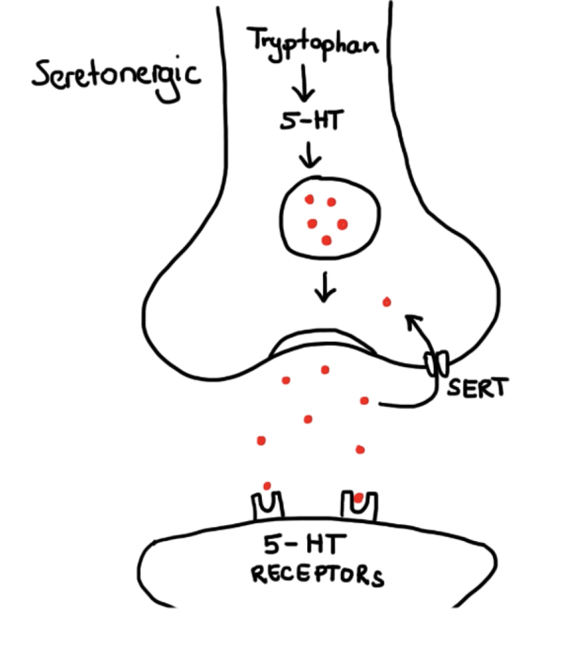
SLC6A1 regulates the reuptake of an important neurotransmitter called gamma-Aminobutyric acid (GABA) which is essential for brain development and working memory. Finding this gene was no simple task and took years. It all started with a critical breakthrough about schizophrenic individuals.

In 2015, a research team at the University of Pittsburgh found that one of the key differences between people with schizophrenia and those without is that schizophrenic individuals have impaired GABA transmission which affects their basic cognitive function. From here, a research team at Cardiff University hypothesized that the impairment is genetic since genetic code is the basis for the creation of neurotransmitters. They rigorously worked through thousands of files of genetic data from the EU biobank of schizophrenic and non-schizophrenic people, cross-referencing each until they found copy-number variants (variations of strings of nucleotides in genetic code) that were common between people with schizophrenia.

Finally, the team performed, “the largest analysis of coding in schizophrenia to date.” They gathered 674 schizophrenic genetic trios (trios composed of two parents with schizophrenia and one child) to analyze the copy-number variants present in their DNA sequences. To their discovery, they found that it was common that within these trios, either one or more had a mutated version of SLC6A1 that impairs GABA transmission. This alters brain development and increases risk to schizophrenia. Being able to narrow down the impairment of GABA in schizophrenic individuals makes this a significant development because it weaves previously known information with new information, cluing researchers in that they are moving in the right direction.

With this major advancement on hand, the next question begs to be asked, where do we go from here?

Robert Cotes, a doctor at Emory Psychiatry, said in an interview with Sanjay Gupta that, “we have made a lot of advancement in the study of schizophrenia and then the etiology and in the neurobiology of schizophrenia. Unfortunately, I don’t know how much of those have really translated to improved outcomes.”

It is devastating that most of the discoveries in schizophrenia research go unfollowed or lead to dead-ends. According to the Treatment Advocacy Center, Schizophrenia affects 1% of the world population and numbers keep rising. In the US alone, approximately 2.6 million people aged 18 and older are have schizophrenia with an estimated 40% of individuals with the disorder going undiagnosed. Schizophrenia is becoming more of a problem now than ever.

There are many directions to go from here. The current focus is on confirming the results from Cardiff University and helping to improve the quality of life of people with schizophrenia. This study is the first of its kind. The team acknowledges in their paper that their sample size is too small to make any large generalizations, but they are hopeful that the discovery of mutated SLC6A1 will help push for more researchers to find other genes/mutations that cause schizophrenic like symptoms. With more studies on specific genes in relation to schizophrenia, research will be able to confirm or deny the current findings, helping to determine if they are truly significant.

The team also hopes to inspire others to start producing new, more effective, and updated medications/treatments that target SLC6A1 to subdue schizophrenic symptoms. This will help ease people with schizophrenia and help them get back to living their normal lives without the worry of having a schizophrenic attack.

Of course, the ultimate goal for any health ailment is to find the cure. With enough time, technology, and research, it will be possible to stop the effects of mutated SLC6A1 completely. For example, a combination of gene therapy with this new knowledge can be used to fix the mutation, essentially creating a possible cure for schizophrenia. If SLC6A1 truly is the cause of  the disorder, it may be completely eradicated in the coming years.

Even if the cure for schizophrenia does not come out of this new study, the cure is on the horizon. There should be a greater push for schizophrenia research and now is not the time to slow down. With more work, it will be possible to help people like Turner and millions to get their lives back from the grasp of this destructive disorder. People with schizophrenia no longer need to feel scared, alone, and trapped. Soon, we can help them become confident, included, and free.

By: V. Palanivel

References: 

Bergen, S. E., Ploner, A., Howrigan, D., O’Donovan, M. C., Smoller, J. W., Sullivan, P. F., et al., (2019). Joint contributions of rare copy number variants and common SNPs to risk for schizophrenia. American Journal of Psychiatry, 176(1), 29-35. doi:10.1176/appi.ajp.2018.17040467 https://ajp.psychiatryonline.org/doi/10.1176/appi.ajp.2018.17040467

Frankle, W. G., Cho, R. Y., Prasad, K. M., Mason, N. S., Paris, J., Himes, M. L., et al., (2015). In vivo measurement of GABA transmission in healthy subjects and schizophrenia patients. American Journal of Psychiatry, 172(11), 1148-1159. doi:10.1176/appi.ajp.2015.14081031 https://ajp.psychiatryonline.org/doi/pdf/10.1176/appi.ajp.2015.14081031

Gupta, Sanjay. (2014). Why Is Schizophrenia So Hard to Treat?. Everyday Health. https://www.everydayhealth.com/hs/schizophrenia-caregiver-guide/sanjay-gupta-why-is-schizophrenia-so-hard-to-treat/

Rees, E., Han, J., Morgan, J., Carrera, N., Escott-Price, V., & Pocklington, A. et al. (2020). De novo mutations identified by exome sequencing implicate rare missense variants in SLC6A1 in schizophrenia. Nature Neuroscience. doi: 10.1038/s41593-019-0565-2 https://www.nature.com/articles/s41593-019-0565-2

Treatment Advocacy Center. (2020). Schizophrenia – Fact Sheet  https://www.treatmentadvocacycenter.org/evidence-and-research/learn-more-about/25-schizophrenia-fact-sheet

Image Credits: 

Stress in Mind, Adobe Stock, https://cdn.mindful.org/StressMind.jpg?q=80&fm=jpg&fit=crop&w=1400&h=875

A pharmacist is advising a patient about his medication.Combatting Disparities in Rural Health Care through Community Pharmacy

If you are suffering from a headache, try this medicine! It’s about time you took back control of your day. Side effects may include fatigue, seizure, or death. Get started on your path to better health with the medicine that gives you your day back!

While most people tune out drug commercials like the one above, the infamous part of the commercial that includes a laundry list of side effects has become almost humorous. The idea of suffering from some of the symptoms listed may seem comical and exaggerated. However, such things are possible without proper supervision.

For populations in rural areas, over the counter medicine or even prescribed medicine is poorly administered. According to an article in American Journal of Health System Pharmacy, “rates of   overdose from prescription opioid misuse are highest in states with  large rural populations.” The laundry list of side effects isn’t that uncommon for populations with high rates of drug misuse.

In fact, a study in the American Journal of Health System Pharmacy mentioned that 1 in 4 Americans live in rural areas. The number of people subject to misuse of prescription medicine is quite high in rural settings due to geographic isolation. Rural areas are characterized as being far outside main cities with small populations and settlements.

Consequently, such areas have reduced access to health care providers. According  to  the  National  Rural   Health   Association, “access to healthcare services, including primary care, specialty care, psychiatry services,  and  hospital  care,  is  often  lacking  in  rural  areas.” Due to patient isolation from their health providers, rural areas have higher rates of health errors. So, what is the role of pharmacists in decreasing adverse effects and drug misuse?

Now, pharmacists are officially licensed as clinical specialists with the power to offer expansive services to patients. However, pharmacists couldn’t always offer such care.

Beginning in the 1920s to 1950, formal pharmacy practice centered around the compounding, the creation of prescription medicine by a pharmacist. In fact, in an article in the Pharmacy journal, this era of pharmacy practice was called the “Soda Fountain era.” Pharmacists just received and filled orders like a Soda Shop. Given only the most basic of tasks, pharmacists across the country were ready to do more!

From the 1950s to the 1970s, pharmacists began completing more tasks related to clinical patient care ,which is the observation and treatment of illnesses. With more time spent in doctoral training, pharmacists gained exposure to a multitude of different cases. For the first time, pharmacists were given the opportunity to demonstrate their potential to diagnose and treat patients, offering comprehensive care to the masses.

Beginning in the 1980s, the pharmacy practice underwent major changes: the degree program was lengthened to six years, pharmacy became an official clinical practice, and the first community pharmacy resident programs began. Pharmacists were officially given their rights to offer clinical care to patients in partnership with physicians. With the licensure of pharmacists as clinical specialists, pharmacists were encouraged to move to rural areas that were in need of primary care and fill the role as physicians. However, while pharmacists were allowed to practice as clinical specialists, they weren’t properly trained for rural practice.

Community pharmacy could help bridge the gap between patients residing in rural areas and pharmacists given the proper training. Therefore, pharmacy programs across the country are implementing comprehensive curriculum which includes residencies in rural sites. Specifically, the University of North Carolina at Chapel Hill’s Eshelman School of Pharmacy offers a rural pharmacy satellite program. As the number one pharmacy school in the country, the move to increase and improve rural pharmacy education demonstrates the future of rural health care.

Cohort studies conducted at other pharmacy school support the worth of working at rural sites. The time spent in an actual community pharmacy effectively exposes students to important practices, improving their confidence in their abilities.  According to the Eshelman School of Pharmacy, through comprehensive training, pharmacists leave school ready to accomplish their goal of improving population health in rural areas.

To improve population health, pharmacists are expected to be the leaders in their community. A community pharmacist is required to complete a multitude of task such as drug management, chronic illness management, and other drug counseling. They must proactively monitor patient’s care plans to ensure proper drug administration.

Telepharmacy is a new video conferencing tool created to improve drug administration and prevent drug misuse. Telepharmacy enables pharmacists and patients to conduct more frequent conversations. The tool can be used to discuss treatment plans and other drug counseling.

Accessibility is the main goal for health professionals as they seek to alleviate disparities in rural health care. With expanding fields such as community pharmacy with tools like telepharmacy at their disposal, community pharmacists are ready to meet the needs of populations without adequate health care.

The next time you are thinking of picking up a random patented headache imagine the laundry list of random symptoms you could develop. Take advantage of the services that your local pharmacist can offer. They won’t leave you astray and you can leave the pharmacy with less pain then you entered with.

By: K. Waite

References:

Hartley, David. “Rural health disparities, population health, and rural culture.” American journal of public health vol. 94,10 (2004),pp. 1675-1678. doi:10.2105/ajph.94.10.1675

Patterson, Brandon J., et al. “Rural Access to Clinical Pharmacy Services.” Journal of the American Pharmacists Association, vol. 54, no. 5, (2014), pp. 518–525.,doi:10.1331/japha.2014.13248.

Scott, Mollie, et al., “Creating a new rural pharmacy workforce: Development and implementation of the Rural Pharmacy Health Initiative”, American Journal of HealthSystem Pharmacy, Vol. 74, Iss. 23,(1 December 2017), pp. 2005–2012,doi.org/10.2146/ajhp160727

Urick, Benjamin Y., and Emily V. Meggs. “Towards a Greater Professional Standing: Evolution of Pharmacy Practice and Education, 1920–2020.” Pharmacy, vol. 7, no. 3, (2019), pp. 1-11. ProQuest, http://libproxy.lib.unc.edu/login?url=https://search.proquest.com/docview/2312274102?accountid=14244, doi:http://dx.doi.org/10.3390/pharmacy7030098.


Image Credits:

Baer, Rhoda,”Man Consults with Pharmacist”, National Cancer Institute Visuals Online, https://visualsonline.cancer.gov/details.cfm?imageid=7517


The Endless Possibilities of Skin Cells

Chandan Sen, a researcher at Ohio State, holding up the Tissue Nanotransfection Device

Chandan Sen, a researcher at Ohio State, holding up the Tissue Nanotransfection Device

What if I told you that you had roughly 35 billion cells that could be reprogrammed to help save your failing kidney, or be altered to help your wounds heal faster and stronger? Due to the newfound ability to transform skin cells to iPSCs (induced pluripotent stem cells), that and much more is now possible. 

The average NFL football stadium has a seating capacity of 69,000 people when fully packed. As of January 2019, more than 113,000 people were documented being on a waiting list for a life-saving organ transplant. That’s almost twice the capacity of the football stadium, and that’s only the documented cases. There could hundreds, or even thousands more just in the United States alone. For years, scientists and researchers have tried to come up with a solution for organ shortages around the world, but to no avail. However, a new organ manufacturing technique involving skin cells has come to light, and it may be the miracle we were all waiting for. 

Skin is a remarkable part of our bodies. Not only is it our largest organ, but it also serves multiple purposes. It protects from outside factors such as sunlight or some light physical damage, helps us feel from the nerves embedded in them, and helps us cool down through the sweat glands. Skin is composed of three parts: the Epidermis, the Dermis, and the subcutaneous layer. The Epidermis is composed of four layers, the top two of which are a cycle of dead and replacement cells. Melanin comes from the last layer of the Epidermis. The Dermis layer is made up of blood vessels and provides the skin’s flexibility. Lastly, the subcutaneous layer is just fatty tissue. 

The discovery of being able to genetically alter skin cells to have them become one of three types of induced pluripotent stem cells (iPSCs), which are just essentially stem cells of the embryo, placenta, or extra-embryonic tissue, is a modern discovery that is fairly well known in the scientific community. But researchers at Ohio state have taken the concept of iPSCs to the next level. They have managed to create a trial involving a device called the Tissue Nanotransfection device (TNT), a noninvasive tool that can alter skin cells with just a touch, and transform them to the intended type of cell, including organ cells. The test subjects for this experiment were a mouse and a pig with injured legs that did not have blood flow. Within a week of applying the Tissue Nanotransfection device, active blood vessels were found in both the test subject’s legs, and within two weeks the legs were saved. But the use of the TNT device goes beyond just organ manufacturing; nerve cells were also formed by transforming skin cells to help mice who had strokes to recover. 

The key to unlocking all of this was the implementation of skin cells into iPSCs. One of the most famous origins of this technology was Dr.Yossi Buganim and his team at The Hebrew University of Jerusalem. Together they found a set of five genes that once inserted into the skin cells, would alter them into the iPSCs cells, the cells that create fetuses. When studying the structure of the skin cells, they found out that once the set of genes is introduced into the skin cells, the cell first “loses” its identity and then gains one the function of one of the three embryonic cells, known as an iPSCs

In the short time that the technology for the iPSCs has been available, modifications to make it safer and more effective has begun. One of the reasons that the induced pluripotent stem cell transformation works is from its use of transcription factors. Transcription factors are proteins that regulate the rate in which DNA is converted to its counterpart RNA. In previous trials, the transcription factors generally used are called Klf4 and c-Myc. Researchers Wenwen Deng, Cao Xia, Chen Jingjing, and Zhang Zhijian replaced those transcription factors with microRNA 302-367, which was chosen out of seven other compositions, and tested in mice with compromised immune systems. After gathering the data, the results showed that the change in transcription factors created a smaller chance of genetic modification but also helped the reprogramming efficiency to go up tremendously. 

But the benefits of iPSCs don’t just stop at organ manufacturing. Scientists from Stanford University turned skin cells into blank iPSC cells and had them grow in monolayers (single layer, sometimes in Petri dishes). After putting them in a dish and seeing colonies form, they added a mixture of compounds as an attempt to try to make them form immature brain cells. Eventually, a formation of neurons and astrocytes (star-shaped cells found in the brain) was found within the dish. Once the scientists cut the cells in half they found a network that is similar to the cortex of the human brain. Upon further testing, they found that 80 percent of the neurons in the cells could fire when stimulated by the scientists and 86 percent contributed in the activity that resembles what is normally found in the brain. A possible outcome of this experiment is being able to attempt to understand the development of the brain and its disorders in a different and closer way, by being able to manipulate its growth and process.

Although fairly early in development, skin cells are becoming much more involved in modern medical research. The abundance, as well as malleability of the cells, could be the key to solving many of the problems that affect our health today, as well as being able to study organs and parts that we couldn’t before. Who knows, maybe the cure to cancer could lie in the palm of our hand. Quite Literally. 

By: S.Singh

References:

Andrew, Elise. “Scientists Grow Mini Brains From Patient Skin Cells.” IFLScience, IFLScience, 11 Mar.2019, https://www.iflscience.com/brain/scientists-grow-mini-brains-patient-skin-cells/

Deng, Wenwen.Xia, Cao, Jingjing, Chen.Zhijian, Zhang “OOPS.” Digital Object Identifier System, 2015, doi.org/10.1021/acsami.5b06768.

“Embryo Stem Cells Created From Skin Cells.” ScienceDaily, ScienceDaily, 2 May 2019, www.sciencedaily.com/releases/2019/05/190502143437.htm.

Geoshen. “NFL Stadiums Ranked by Seating Capacity.” NFL Stadiums Ranked by Seating Capacity, geoshen.com/posts/nfl-stadiums-ranked-by-seating-capacity.

Ohio State University. “Ohio State Researchers Develop Regenerative Medicine Breakthrough.” Ohio State Researchers Develop Regenerative Medicine Breakthrough, The Ohio State University, 12 July 2018, news.osu.edu/ohio-state-researchers-develop-regenerative-medicine-breakthrough/.

“Organ Donation: Facts vs. Fiction.” UCI Health, UCI Health, 23 Apr. 2019, www.ucihealth.org/blog/2019/04/organ-donation.

“Transplant Trends.” UNOS, unos.org/data/transplant-trends/.

Image Credits:

The Ohio State University Wexner Medical Center, Researcher Chandan Sen with the nanotechnology-based chip designed to deliver biological “cargo” for cell conversion, Ohio State News, https://news.osu.edu/ohio-state-researchers-develop-regenerative-medicine-breakthrough/

The Measles Epidemic is Surging


Baby with Measles

Baby with measles in Vienna, Virginia

“The scare tactics worked with the Walthers, and they decided not to immunize their daughter. It was a choice they lived to regret,” said Betty Bumpers. “Mary Catherine was lucky. She survived, but her ordeal certainly prompted her parents to question the health information they found on the Internet.”

Mary Catherine was born in the United States. As an infant, her parents decided not to give her the recommended childhood vaccinations. Little did they know, their baby girl would become deathly ill before turning the age of one, and her sickness could have been prevented by a simple vaccination. 

Vaccinations were invented in the late 1700s as a way to protect against deadly diseases. Edward Jenner invented the first vaccine against smallpox. He noticed that milkmaids who were exposed to cowpox did not develop smallpox. He then used the material from inside cowpox to make a vaccine. Over the next two hundred years, many vaccines were developed and scientists continue to discover new vaccines against emerging infections. Vaccines now exist against polio, measles, mumps, rubella, chickenpox, and meningitis and are part of routine childhood doctor visits.

Vaccinations prevent two to three million deaths each year, according to World Health Organization. In a 2018 report, Centers for Disease Control (CDC) reported there were more than 140,000 deaths worldwide due to measles. Since the release of the CDC report, the number of reported measles cases jumped 300% worldwide within the first three months of 2019 compared to the first three months of 2018. The majority of these deaths occurred in low-income countries due to lack of access to the measles vaccine but in high-income countries, like the United States, measles has started to make a comeback due to the rise of the anti-vaccination movement. 

The Anti-Vaccine Movement started in Europe and the U.S during the mid-1800s. Since this movement began, it has progressively grown over the past 200 years with an increasing number of parents either delaying or refusing altogether to let their infants receive vaccinations. The most common reason for parents to refuse vaccines for their children is due to religious beliefs and fear of side effects. These views, for the most part, are guided by misinformation and false science.

One of the major reasons parents refuse to allow their children to have vaccines is fear of autism, specifically related to the measles, mumps, and rubella vaccine (MMR). In 1998, Andrew Wakefield fueled this fear by publishing a study in the British medical journal, The Lancet. He claimed that infants who received the MMR were more likely to develop autism. His studies were filled with fraudulent research and false information and The Lancet later went on to remove his publication from their journal. Unfortunately, the damage had been done: negative propaganda exploded around the MMR. The number of vaccinations plummeted and the incidence of measles cases increased

In response to the misinformation around the MMR and autism, Dr. William Schaffner, Professor at Vanderbilt, compared rates of autism in children who were vaccinated and those who were not, and he found that the rates of autism were not increased among those children who had received the vaccine. He concluded,

“MMR vaccination does not increase the risk for autism, does not trigger autism in susceptible children, and is not associated with clustering of autism cases after vaccination.”

This study helped debunk the misinformation that linked autism to measles. 

In the fall of 2018, one of the largest US measles outbreak occurred among New York’s Orthodox Jewish Community. More than 285 under-vaccinated infants were infected in three different communities and this outbreak prompted health officials to give out mandatory immunizations. It happened because the NY Orthodox Jewish community is a very insular group. There have been many anti-vaccine campaigners in New York who were giving false information with regards to the measles vaccinations. These campaigners were spreading the message that vaccines were not safe or effective, which is completely incorrect. 

Parents who refuse vaccines think they are actually protecting their children, but in reality, they are placing them at risk of getting preventable diseases. In addition, these children do not have a voice or a choice. Refusing a child a vaccine is selfish because it affects that child, but also the community. As well as protecting the individual who gets a vaccine, vaccinations help keep those who have not been vaccinated safe, this is called Herd Immunity. Many diseases could be almost eradicated with the widespread use of vaccines. 

Robert Pearl MD, Stanford professor, former medical group CEO, and forbes.com contributor said it best in regards to why we should be using vaccinations:

“When my father was a child, his sister died of measles. Her death stayed with him throughout his life. That was before we had a vaccine to prevent measles. If she had been born in the 21st century, she might not have died at age 6… We need to ask ourselves this:  If economics are not the deterrent, why would a parent put their child’s life at risk by voluntarily foregoing a life-saving preventive measure? The answer is a combination of false science, outdated anecdotes, and fear-mongering.”

Although vaccinations are still voluntary, they hold an immense amount of importance to communities worldwide. All parents want to do the right thing and protect their children, but anti-vaxxers are instead placing their children at risk. Ongoing education is really important to promote uptake of childhood vaccines. The Centers for Disease Control Prevention has an obligation to help monitor outbreaks of preventable diseases and to work with local communities to disseminate information and dispel false rumors about vaccinations. Getting anti-vaccine parents to change their minds is difficult, and education is key. 

By: W Stringer

References:

  1. Belluz, Julia. “New York’s Orthodox Jewish Community Is Battling Measles Outbreaks. Vaccine Deniers Are to Blame.” Vox, Vox, 10 Apr. 2019, www.vox.com/science-and-health/2018/11/9/18068036/measles-new-york-orthodox-jewish-community-vaccines.
  2. Bumpers, Betty. “Unprotected People Reports: General.” Some Parents Fall for Vaccination Scare Stories, with Deadly Results, 1 Jan. 2001, www.immunize.org/reports/report038.asp.
  3. Hoffman, Jan. “How Anti-Vaccine Sentiment Took Hold in the United States.” The New York Times, The New York Times, 23 Sept. 2019, www.nytimes.com/2019/09/23/health/anti-vaccination-movement-us.html
  4. Iannelli, Vincent. “History of the Anti-Vaccine Movement.” Verywell Health, Verywell Health, 24 Jun. 2019, www.verywellhealth.com/history-anti-vaccine-movement-4054321.
  5. Koenig, Debbie. “2019 Measles Outbreak: What You Should Know.” WebMD, WebMD, 11 Apr. 2019,  www.webmd.com/children/news/20190411/2019-measles-outbreak-what-you-should-know.
  6. Pearl, Robert. “A Doctor’s Take On The Anti-Vaccine Movement.” Forbes, Robert Pearl, M.D, 20 Mar. 2014, www.forbes.com/sites/robertpearl/2014/03/20/a-doctors-take-on-the-anti-vaccine-movement/#3b914e3328dc
  7. Shimizu, Yoshi. “Measles.” World Health Organization, World Health Organization, 5 Dec. 2019, www.who.int/news-room/fact-sheets/detail/measles.
  8. William Schaffner, “Anti Vaccination Movement Fuels Return of Measles.” Hospital Employee Health, 38. 5 (2019), 

Image Credit:

Lowell Georgia, Baby with measles in Vienna, Virginia, Baby Center, https://www.sciencesource.com/archive/Measles-SS2152242.html


Treat Seasonal Affective Disorder with Light


The Mind-Body Problem by ROZ CHAST

“Hurry, get up.”

You stare at the ceiling and murmur to yourself. Your head is splitting, your body feels heavy, and on this cold, bleak winter morning, once again, it feels too difficult to get out of the bed. Even when you finally make this great achievement just like crossing the finish line, you still feel exhausted, sluggish, and hard to focus on work during the winter’s day.

But don’t blame yourself for this. Feeling more tired and depressed in winter is not your fault. Instead of being simply explained by declining willpower in cold weather, this phenomenon actually arises from a series of physiological effects called Seasonal Affective Disorder, which is experienced by more than 10 million people in this country.

The mechanism behind Seasonal Affective Disorder is complicated. As a carbon-based creature, your feelings of happiness and well-being are built on numerous reactions between organic molecules. One indispensable regulator is serotonin, a transporter of neural signals, which is synthesized in presynaptic neurons and released to the synaptic cleft, then absorbed by serotonin receptors. This harmonious co-ordination makes you feel positive after having a chocolate bar or finishing your lab report.

However, based on cross-sectional neuroimaging studies, Brenda McMahon and his research group found that in winter, people with Seasonal Affective Disorder have 5% more serotonin transmitters (also called SERT), which will “capture” free serotonin molecules and transport them back to presynaptic neurons.

Screenshot of a YouTube video course

Signaling in Serotonergic Synapses by Speed Pharmacology

The direct treatment for this is taking pills designed to inhibit the concentration of serotonin transporter. The most popular choice is SSRI, i.e. Selective Serotonin Reuptake Inhibitors, including Prozac, Paroxetine, etc. By binding to and inactivating serotonin transporters, this drug increases the level of serotonin available in the synaptic cleft. As one of the most prescribed drugs in America, to some individuals, SSRI works well.

But there are two problems. Firstly, SSRI has nonnegligible side effects. In an experiment conducted by Kylie Thaler, up to 27% of Seasonal Affective Disorder patients treated with SSRI had adverse effects such as nausea and headache and finally dropped out of the research. Additionally, overall, this drug only works about 40% of the time.

Scientists then turn to an indirect method to fortify the level of serotonin in winter: synthesizing vitamin D, which is an essential micronutrient in helping maintaining an optimal environment to synthesize serotonin, and for human beings, exposure to sunlight provides over 90% of the daily dosage of vitamin D. As a result, it’s easy to understand why we may suffer from vitamin D deficiency and unbalanced extracellular fluid serotonin concentration during the dim, gloomy winter, and it seems like sun tanning becomes the go-to strategy of Season Affective Disorder.

But as you may have noticed, the key of enhancing vitamin D synthesis is exposing your skin directly to the sunlight without any protection from sunscreen or sunglasses to filter the light in ultraviolet region (UV), only which, however, functions in turning steroids into vitamin D. Since UV light will trigger mutations in gene sequence and destroy our genetic information, it’s not a good deal to treat Seasonal Affective Disorder at the price of skin cancer.

Till now, it seems like we reach an impasse. But as mentioned before, besides serotonin, there are many other organic chemicals participating in the formation of Seasonal Depressed Feelings. One main character is melatonin.

As a pivotal monitor of your biological clock, melatonin to your circadian rhythms is just like Caesar to Rome. It designs your in-body calendar, regulates your circadian phase, and makes a balance between sleep and awake time.

When the dark signal is detected by the retina, the pineal gland is stimulated to produce melatonin while the melatonin production is inhibited when the light signal is detected.

Melatonin Helps Maintain the Circadian Rhythm by arigo Biolaboratories (revised)

The way that our body uses melatonin to control our biorhythms is simple: when light is conveyed by the retina, opsins, a group of proteins, turns the light signal into a neural signal, which reaches the suprachiasmatic nucleus in your hypothalamus, and suppresses the melatonin release inside pineal gland. In 2017, Ashutosh Jnawali proves that melatonin release is totally dependent on light exposure which could effectively decrease the activity of a gene called AANAT, which catalyzes the penultimate step in the synthesis of melatonin. Hence during winter, the decreased daytime triggers a higher level of melatonin, and we are more likely to feel like a bear ready for hibernation (just as bears eat a lot to store energy before hibernation, don’t feel surprised at your unusual good appetite in winter, which is another symptom of Seasonal Affective Disorder).

Given this mechanism, a non-drug treatment based on your natural physiological function is Light Therapy. The rationale of this strategy is to suppress the daytime melatonin synthesis, improves your sleeping quality, and decrease Seasonal Affective Disorder symptoms by exposing to extra light in the relatively dimmer season.

In 2019, Pierre A. Geoffroy found that in clinical settings, compared to the combination of antidepressant drugs and placebo, there is a superiority of Light Therapy + Placebo (54% remission rate) in relieving the symptom of both seasonal and non-seasonal depression (50% remission rate). Additionally, the effect of a combination of Light Therapy + antidepressant drugs is confirmed to be considerably stronger than drugs + placebo, which further proves the therapeutic function of light in Seasonal Affective Disorder.

Here, you may start to wonder: given the same exposure to light, why does the process of synthesizing vitamin D come along with skin cancer risk, but Light Therapy has nothing to do with it? The reason is in Light Therapy, the light we make use of (i.e. the signal captured by your retina) is visible light rather than ultraviolet light. Put it in another way, if you want to get good use of sunlight to control the melatonin synthesis, feel free to use sunscreen to protect your skin.

Of course, for those who live in the place heavy on raining days in winter, a better choice is light box, which is designed to filter the UV light and keep only visible light to treat Seasonal Affective Disorder.

To the patient with eye problems such as glaucoma, light stimulation to eyes could be dangerous. But this doesn’t mean that light therapy is inaccessible to them: A pilot study conducted by Markku Timonen found out that to regulate the secretion of melatonin, light can be transmitted via ear canals, i.e. through non-opsin mediated pathways.

Valkee Human Charger “Is Amazing” by Valkee

Though over species, most of the light signal is transduced by opsin in the retina, there is a special extra-retinal protein, called OPN3, which can also converse photon into neural responses. In another experiment conducted by Heidi Jurvelin, this extravisual transduction is proved to be effective as 92% of patients with Seasonal Affective Disorder felt better after receiving lumen bright light daily into both ear canals for 4 weeks.

Overall, no matter through eyes or ears, Light Therapy could be a good choice to overcome the depressed feelings during winter with the benefits such as low cost, few side-effects, etc.

I Think He’s About to Present Us with an Idea by Frank Cotham

The next time when you feel guilty about the tiredness and low-efficiency in winter, please don’t beat yourself up. Just choose a light box (or a fancy bright light headset, as you wish), open the window, or put on shoes to go for a walk under the sun. Winter is not meant to be a sad season: you can make it warm, bright, and vibrant… as long as you embrace the light.

By: B. Tan

References

Geoffroy, P. A., Schroder, C. M., Reynaud, E., & Bourgin, P. (2019). Efficacy of light therapy versus antidepressant drugs, and of the combination versus monotherapy, in major depressive episodes: A systematic review and meta-analysis. Sleep Medicine Reviews, 48, 101213. doi: https://doi.org/10.1016/j.smrv.2019.101213

Jnawali, A., Backus, B. T., Quinlan, E. M., Llerena-Law, C., Viswanathan, S., Joshi, N., … Ostrin, L. A. (2017, June 23). Physiological Effects of Ten Days of Total Darkness in Humans. Retrieved from http://iovs.arvojournals.org/article.aspx?articleid=2641280

Jurvelin, H., Jurvelin, H., Takala, T., Takala, T., Nissilä, J., Nissilä, J., . . . Räsänen, P. (2014). Transcranial bright light treatment via the ear canals in seasonal affective disorder: A randomized, double-blind dose-response study. BMC Psychiatry, 14(1), 288. doi: https://doi.org/10.1186/s12888-014-0288-6

Mc Mahon, B., Andersen, S. B., Madsen, M. K., Hjordt, L. V., Hageman, I., Dam, H., . . . Knudsen, G. M. (2016). Seasonal difference in brain serotonin transporter binding predicts symptom severity in patients with seasonal affective disorder. Brain, 139(5), 1605-1614. doi: https://doi.org/10.1093/brain/aww043

Melrose, S. (2015). Seasonal affective disorder: An overview of assessment and treatment approaches. Depression Research and Treatment, 2015, 178564-6. doi: https://doi.org/10.1155/2015/178564

Ostrin, L. A. (2019). Ocular and systemic melatonin and the influence of light exposure: Melatonin and light exposure. Clinical and Experimental Optometry, 102(2), 99-108. doi: https://doi.org/10.1111/cxo.12824

Thaler, K., Delivuk, M., Chapman, A., Gaynes, B. N., Kaminski, A., & Gartlehner, G. (2011). Second-generation antidepressants for seasonal affective disorder. Cochrane Database of Systematic Reviews (Online), 12(12), CD008591. doi: https://doi.org/10.1002/14651858.CD008591.pub2

Timonen, M., Nissilä, J., Liettu, A., Jokelainen, J., Jurvelin, H., Aunio, A., . . . Takala, T. (2012). Can transcranial brain-targeted bright light treatment via ear canals be effective in relieving symptoms in seasonal affective disorder? – A pilot study. Medical Hypotheses, 78(4), 511-515. doi: https://doi.org/10.1016/j.mehy.2012.01.019

Tyrer, A. E., Levitan, R. D., Houle, S., Wilson, A. A., Nobrega, J. N., & Meyer, J. H. (2015). P.1.i.048 seasonal variation in serotonin transporter binding in seasonal affective disorder and health: A [11 C]DASB positron emission tomography study. European Neuropsychopharmacology, 25, S328-S328. doi: https://doi.org/10.1016/S0924-977X(15)30406-5

Image Credits

Image 1: “The Mind-Body Problem”, The New Yorker, https://www.newyorker.com/cartoon/a20819

Image 2: “Pharmacology – ANTIDEPRESSANTS – SSRIs, SNRIs, TCAs, MAOIs, Lithium”, Speed Pharmacology, https://www.youtube.com/watch?v=T25jvLC6X0w&t=256s

Image 3: “Melatonin”, arigo Biolaboratories, https://www.arigobio.com/news/besides-for-human-arigo-s-melatonin-elisa-kit-also-works-for-arabidopsis

Image 4: “Valkee Human Charger Is Amazing”, Natural Health and Wellness Light Therapy Products, http://www.lighttherapyproductsblog.com/valkee-human-charger-is-amazing/

Image 5: “I Think He’s About to Present Us with an Idea”, The New Yorker,  https://www.newyorker.com/cartoon/a17588

Trikafta: Breathing New Life Into the CF Fight


Most teenagers do not typically face the risk of death when interacting with others on a daily basis. However, this is the harsh reality for nineteen-year-old Ally Nichting. When she was four months old, Ally’s parents took her to Duke Hospital after they had consistently noticed abnormal digestive activity. It was there that doctors delivered the life-changing news and diagnosed Ally with cystic fibrosis.

Cystic fibrosis (CF) is the mutation of the cystic fibrosis transmembrane-conductance regulator (CFTR) protein, which is located on the surface of the cell membrane. The CFTR protein allows for the passage of chloride in and out of the cell. However, because CF patients have mutated CFTR proteins, they end up with a thick build-up of mucus in their lungs, which is just one, and arguably the most prominent, of the many issues associated with cystic fibrosis.

The dense mucus that builds up in the lungs of CF patients causes them to be more prone to bacteria that grows very easily in that environment, leading to infections in the lungs. CF patients must be very cautious about interacting with one another due to the risk of cross contamination. If two CF patients interact in person, it increases the risk of developing infections because of the different bacteria in each person’s lungs. In most cases, this could potentially cause long-term, severe illness or death. The Cystic Fibrosis Foundation recommends that individuals with the disease stay at least 6 feet away from each other to reduce the risk of exposure to germs that become airborne with a cough or sneeze. Even a simple seasonal cold can send CF patients to the hospital for two weeks or more. 

In addition to build-up in the lungs, mucus can also build-up in other areas, such as the pancreas. The pancreas is important for the release of enzymes that break down food. According to the Cystic Fibrosis Foundation, nearly 90 percent of CF patients experience mucous blockages in their pancreas, thus creating a need for enzyme supplements, to allow food to be properly digested.

Pancreas sells secreting abnormal amounts of mucus

The majority of CF patients end up having mutations within their already mutated CFTR protein. The most common mutation being the F508del mutation, which is the deletion of the 508th amino acid in their CFTR protein sequence. Some patients, like Ally, have more than one mutation, which prohibits her from taking certain medications to treat her various symptoms.

Thankfully, in the past four months, the FDA approved a new cystic fibrosis triple drug treatment, Trikafta, that works for 90% of patients with cystic fibrosis gene mutations, including Ally’s specific mutation. When she got the news, Ally was elated to find out that there was a drug that accommodates patients like her. “A few years ago, a big drug came out that was very promising but it was only able to help certain mutations, and I had one of those mutations but the other one I have is extremely rare so I wasn’t able to be on it. Seeing that this drug helps 90 percent of the CF gene mutation thrills me!” said Ally.

Trikafta is administered orally via pill and delivers three drugs: tezacaftor, ivacaftor, and elexacaftor. Tezacaftor and elexacaftor correct the CFTR protein so they are able to achieve proper placement on the cell membrane’s surface. Ivacaftor is a potentiator that facilitates the opening of the CFTR protein to allow chloride to pass through. 

In a clinical trial comparing Trikafta (tezacaftor, elexacaftor, ivacaftor) to a double drug treatment Semdeko (tezacaftor, ivacaftor), patients were seen to have more clinically robust benefits with the triple drug treatment versus the double drug treatment. Patients on the double drug treatment had a 10-percentage point increase on their pulmonary functions test (PFTs), specifically on the forced expiratory volume in one second (FEV1), whereas patients on the triple drug treatment had a 17.4-percentage point increase in their FEV1.

In addition to increased FEV1 function, doctors also reported that patients had improved sweat chloride concentrations, showing that chloride was able to pass through the CFTR proteins more efficiently. Patients also discovered improved health related quality of life (HRQoL) on Trikafta versus the double drug treatment.

Because there are only about 30,000 cases of cystic fibrosis in the United States, the disease often goes unnoticed or people don’t realize the full extent to which cystic fibrosis affects everyday life, whether it be being able to breathe or just digest food.  “CF affects literally everything, more than people realize. Sometimes I forget the amount it affects,” said Ally. Something as simple as the human touch is taken for granted by so many humans. If Ally comes into contact with the wrong person’s bacteria, she could face extreme health consequences or even death, which is hard to think about as a nineteen-year-old. 

The development of Trikafta is important because it is able to treat a broad range of cystic fibrosis symptoms to an extent that has not been treatable previously. Without awareness, fundraising, and financial support, the development of Trikafta would not have been possible and Ally would not have this new weapon to fight her battle with Cystic Fibrosis. 

S. Ung

References:

Heijerman, Harry G M, et al. “Efficacy and Safety of the Elexacaftor plus Tezacaftor plus Ivacaftor Combination Regimen in People with Cystic Fibrosis Homozygous for the F508del Mutation: a Double-Blind, Randomized, Phase 3 Trial.” The Lancet, vol. 394, no. 10212, Nov. 2019, pp. 1940–1948., doi:10.1016/s0140-6736(19)32597-8.

Kwong, Eugenie, et al. “The Impact of Cystic Fibrosis-Related Diabetes on Health-Related Quality of Life.” Journal of Cystic Fibrosis, vol. 18, no. 5, Sept. 2019, pp. 734–736., doi:10.1016/j.jcf.2019.03.007.

Mahase, Elisabeth. “Cystic Fibrosis: Triple Therapy Shows Promising Results.” Bmj, 2019, p. l6347., doi:10.1136/bmj.l6347.

N/a. “Cystic Fibrosis Foundation.” Cystic Fibrosis Foundation, 0AD, www.cff.org/.

Voelker, Rebecca. “Patients With Cystic Fibrosis Have New Triple-Drug Combination.” Jama, vol. 322, no. 21, 2019, p. 2068., doi:10.1001/jama.2019.19351.

Image Credits:

Lungs: https://www.yourgenome.org/sites/default/files/styles/banner/public/images/banners/facts/fact-cystic_fibrosis_0.jpg?itok=OXqP0uFK

Microscope pancreas image: http://www.pathology.washington.edu/content/page-img/359/spa-img0034__small.jpg


Mice and Men: Why Rodents are Costing Us $100,000,000s in Failed Research Every Year

Black and White Mouse

Black and White Mouse

As I walked in I was in awe, I imagined a research lab to be filled with beakers and microscopes with scientists walking around in lab coats but it wasn’t any of those things. But none of that is what caught my attention, instead, it was the back wall lined with 5 shelves, each shelf held 8-12 cages of mice, something close to 50 in total! 

While they might look cute and harmless, I would eventually learn that those tiny creatures were actually costing the US hundreds of millions of dollars every year in wasted research.

In recent decades there has been an increasing problem with reproducing results in humans from research done on rodents. The hundreds of millions being spent on this research produces no useful results in the end and is essentially wasted.

The wall filled with cages upon cages of mice isn’t an unfamiliar one in a biomedical lab. According to the National Association for Biomedical Research, 95% of the animals used by biomedical researchers are rodents, which translates to roughly 20,000,000 rodents being used annually for animal models in the United States alone! One would assume that if they are being used this often, they must be a very accurate model; however, after doing some research, to my surprise, the answer is no. 

The truth is that this is how it’s always been done and once people are set in their ways, it’s hard to change it.

The Animal Welfare Act of 1966, the only federal law in the United States that regulates the treatment of animals in research, doesn’t cover birds or rodents that were bred for the specific purpose of research. This basically means we can and have done anything and everything to mice in the quest for knowledge. We’ve developed technology to be able to modify mice’s genome to add or remove anything we want. We can make mice that glow in the dark, ones without an immune system, ones that have specific diseases or ones with tumors and the list goes on and on. 

Mice are very unique in the number of different options it gives researchers, but the problem is that the conclusions made from mice research don’t translate well into humans; after all, that is the end goal.

Mice share 97% of the same DNA as humans – this was one of the original reasons why mice were chosen as models for human biomedical research. However, we are starting to realize that 3% makes a whole lot of difference. Dr. Perlman compared the metabolic rates of humans and mice and found that a 30g mouse has a metabolic rate 7 times that of a 70kg human! Our metabolisms have a huge impact on how our bodies respond to different drug treatments and such a drastic difference between humans and mice leads to drastic differences as to the effectiveness of drugs.

Dr. Seok, a researcher studying human inflammatory disease, found that prior to 2013, 150 clinical trials had been conducted looking at different drugs to prevent inflammatory response in humans and none were found to be successful, even though all had passed animal testing.   

The problem has surpassed simply a waste of money and time; it has begun to threaten the lives of humans. 

In an extreme case, Dr. Attarwala reviewed a clinical trial conducted in 2006 looking at a new drug that showed promising results in animal models of increasing antibody function. The drug could have been used in the treatment of autoimmune diseases, a class of diseases in which one’s own immune system attacks itself. The drug was tested on mice and was found to be effective and showed no signs of adverse effects. Once the drug got to human clinical trials, a dose of the drug 1/500 of the amount given to mice was given to 6 volunteers. However, unlike the mice within a few hours, the volunteers were all in multi-organ failure and had to be rushed to the hospital. 

Most people would think that obviously the researchers had messed something up or done this intentionally, but after the incident, the United Kingdom’s Medicines and Healthcare Products Regulatory Agency led an investigation into the clinical trial’s procedures and ethics and found no flaws or malicious intent. The problem was that mice and humans just aren’t the same and we can’t be expected to react the same to medications either. The drug should have been tested on chimpanzees or another animal who’s reaction would have been similar to that of humans rather than rodents.   

Well, if we know there’s a problem, then why don’t we stop using mice? This is a tricky position that many researchers find themselves in nowadays. Many know that using mice for their research might make their findings irrelevant or flawed, especially when trying to make conclusions about humans. On the other hand, many researchers have been using mice their entire lives and it’s difficult to switch to other animal models when your expertise is already in mice. Since the majority of the scientific community also tests on mice, they can compare finding something that isn’t possible across species. 

However, the tipping factor comes down to money. Not only are mice cheaper to buy as well as to maintain, many times researchers aren’t able to obtain grants in order to fund their research unless the research is on mice. So if the research isn’t done on mice, it might not ever get done at all. Imagine how much knowledge we’ve lost out simply because we are afraid to branch out and try something new, even if it was more expensive. Some time ago research funded by the government is what leads to innovations that have since brought us: smartphones, GPS, supercomputers, Google Search Engine, MRIs, lactose-free milk, etc. things we now take for granted, but none would have been possible if at the time people were more concerned with saving money over innovation.

In the search for new medicines, the solution is as simple as funding better projects, even if it means funding less, quality or quantity. We know that what we are doing now isn’t working – we haven’t seen breakthroughs in discovering new treatments in decades. According to the CDC, our most effective treatment for Tuberculosis, a bacterial infection of the lungs, is Isonicotinic acid hydrazide, a treatment that we have been using since 1945. Who knows – maybe one of the projects that weren’t funded because testing on chimpanzees was too expensive might have been the one that discovered a cure for cancer.

By: K Patel

References

Attarwala, H. “TGN1412: from discovery to disaster.” Journal of Young Pharmacists 2.3 (2010): 332-336, https://www.sciencedirect.com/science/article/abs/pii/S0975148310230248.

“Treatment for TB Disease.” Centers for Disease Control and Prevention, 5 Apr. 2016, www.cdc.gov/tb/topic/treatment/tbdisease.htm.

“Laboratory Animals.” NABR, www.nabr.org/biomedical-research/laboratory-animals/species-in-research/.

Perlman, Robert L. “Mouse models of human disease “An evolutionary perspective.” Evolution, medicine, and public health 2016.1 (2016): 170-176, https://academic.oup.com/emph/article/2016/1/170/2802638.

Seok, Junhee, et al. “Genomic responses in mouse models poorly mimic human inflammatory diseases.” Proceedings of the National Academy of Sciences 110.9 (2013): 3507-3512, https://www.ncbi.nlm.nih.gov/pmc/articles/PMC3587220/?tool=pmcentrez&report=abstract.

Image Credit

Black and White Mouse 51340, Pexels, https://www.pexels.com/photo/black-and-white-mouse-51340/

Forgetting Fears of Fertility: How Modern Fibroid Treatments Raise Chances for Children

cartoon uterus with submucosal fibroids

Uterine Fibroids

About 70% of women will have fibroids at some point in their lives, according to Guido. If this is such a common occurrence, why then are the treatment options so scarce and generally debilitating towards the patient, and why do so few people even know what fibroids are? Many women get fibroids, which are noncancerous tumors that grow within the uterus that do not on their own affect fertility, but traditional treatment leave women infertile.However, there are new treatments that can both treat these tumors while leaving the woman’s fertility unscathed and giving her a fast recovery.

Traditionally, Guido claims the main treatment option for fibroids is to completely remove their source, the uterus, in a hysterectomy procedure . While this does eliminate the possibility of recurrence of fibroids and relieves all symptoms caused by them, by removing the uterus, women are left infertile. This procedure is also extremely invasive and can leave patients with long recovery periods. Leaving the fibroids be is also not an option, even if they are asymptomatic, as they can cause serious issues with pregnancy. Young states that mortality for mothers can range from 25-50% and mortality for the baby is even higher for pregnancies in a fibroid uterus. Many obstetricians will terminate the pregnancy rather than risk the mother carrying to full term. What are prospective mothers to do, should they have fibroids then?

Thanks to advancements in technology, there are some new treatment options for women that hope to maintain fertility and manage their fibroids. One of these options, as explained by Guido, is radiofrequency ablation, which is a technique that uses alternating currents to heat up and kill the tissue of the fibroids. While there is currently only one procedure that is approved by the FDA, called the Acessa procedure, it has shown huge improvements in the lives of many women treated, with their personal evaluation of quality of life improving by an average 82%. Most patients are also released on the same day of the procedure, and due to it being minimally invasive, recovery time is a lot shorter when compared to a hysterectomy. However, there are some downsides, such as this being a new treatment and the studies being very limited in their population sizes. There is also the possibility that further treatment will be needed, according to Guido, should the Acessa procedure not relieve the symptoms, and these further treatments may include an eventual hysterectomy. However, compared to the traditional methods of treatment for fibroids, radiofrequency ablation offers the best chance at relieving symptoms while maintaining fertility.

Another highly effective treatment option while being non-invasive is MR-guided High-Frequency Ultrasound, which uses a beam of ultrasound light to heat up the fibroid tissue and kill it. Siskin states that this procedure requires no incisions and effectively uses MRI to map out the treatment area. This method, although again only studied on a few patients, has the benefit of a quick recovery due to being so minimally invasive. However, according to both Siskin and Guido, the main drawback of this procedure is that it can be easily affected by other issues in the body, such as the bladder or bowel obstructing the ultrasound beam. Should these be present in the patient, this procedure cannot be done at all, leaving this treatment limited in the number of patients it can be helpful towards. Another limitation is that it cannot be used to treat multiple fibroids at once. Although Siskin posits that multiple treatments are theoretically possible but not yet studied. Those that can have the procedure are shown to have their fibroids reduced in size and have minimal pain caused by the procedure, leaving High-Frequency Ultrasound still a good technique to be both minimally invasive and fertility preserving.

Another new treatment is in the form of a drug known as Uliprisal acetate which has been shown in clinical trails to help relieve the symptom of excessive bleeding and reduce fibroid size. As the study  by McVeigh states, “many patients [have] anaemia, which complicates surgical treatment options”. In a very credible study, being that it was double-blind and a large population size, Uliprisal acetate was shown to be effective in small doses, although larger doses were shown to have some negative side effects. While this drug is not on its own a complete treatment for fibroids, it can help to lessen the symptoms greatly and for long periods while the patient awaits surgical intervention. However, McVeigh notes that this drug is limited in its capabilities, not being effective on larger fibroids and being only useful to relieve symptoms pre-surgery. Still, being able to manage symptoms before more involved measure can be taken, can greatly improve the quality of life for women with fibroids and may prompt doctors to take less serious surgical measures.

With all these treatment options, women affected by fibroids during their reproductive years certainly have options to maintain their fertility while managing their symptoms. While no procedure will ever be as completely effective in removing the source of the problem as a hysterectomy, treatments such as radiofrequency ablation, high-frequency ultrasound, and drug therapies can all be used to greatly reduce fibroids and their symptoms. These modern procedures have made maintaining fertility and being able to quickly recover from treatments no longer fantasy and the many women who will be affected by fibroids at some point in their lives no longer have to rely on the harsh traditional methods.

By: A Vigil

References

Guido, R., M.D., & Stuparich, M., M.D. (2016). Radiofrequency ablation: New paradigm for treatment of fibroids. Contemporary OB/GYN, 61(10), 12-18. Retrieved from http://libproxy.lib.unc.edu/login?url=https://search.proquest.com/docview/1837164184?accountid=14244

McVeigh, E. (2012), Ulipristal acetate: new oral treatment for uterine fibroids. Prescriber, 23: 17-22. doi:1002/psb.981

Siskin, G. (2006). New Treatments in Uterine Fibroids. Techniques in Vascular and Interventional Radiology, 9(1), 12–18. doi: 10.1053/j.tvir.2006.08.004.

Young. (2007). The treatment of uterine fibroids. The New Zealand Medical Journal (Online), 120(1258) Retrieved from http://libproxy.lib.unc.edu/login?url=https://search.proquest.com/docview/1034235948?accountid=14244

Image Credits

episy2, fibrome uterin, pixabay, https://pixabay.com/illustrations/fibroids-fibroids-model-2947712/


Music Powers

Music makes you smarter. It’s like a super vitamin for you brain. Research has proved that listening to music gives it an exercise but being musically trained gives the brain a whole upgrade. Your body also gets some status buffs too, not to mention the ability to play an instrument and to sing more in tune. Overall, it just makes life somewhat easier in the long run.

Music is something all of us have experienced. As children our parents would sing lullabies to sleep, or we would listen to the radio in the car, playing the same “hit” songs that have played everywhere for the past month. Even for those who are hard of hearing, music can be experienced through vibrations where the rhythm of music can be felt. In a sense, music is everywhere. The sound of speech has tone, pitch, timbre (the character of the sound that distinguishes certain instruments from others), and rhythm. We listen to these sounds so often that we don’t pay attention to these details, much less even think about them. As easy as it seems, our brains are constantly splitting the sounds into their most basic forms, sound waves, analyzing them, then putting them back together to deliver to us what we perceive as music.

Every time we listen to music, or anything like it, we exercise our brains. Specifically, our right brain, which controls creativity, intuition, and artistic ability.Though just listening to music exercises our brains, learning how to make music builds the brain even more. It connects the two hemispheres of the brain by building more neurons between them, greatly strengthening the corpus callosum (the part of the brain that connects the two hemispheres). This allows for the two sides to send signals to each other with more efficiency, and less time. This boosts creativity, motor coordination, and general processing speeds. According to research done by The National Institutes of Health and John F. Kennedy Center for the Performing Arts , children who are learning the art of music have better language skills, attention span, visuospatial perception, and executive functions than that of those who have no musical inclination. Those who have gone under musical training early in life also experience a long-term anxiolytic effect, which means that they are more resistant to anxiety, thus improving emotional health and focus, and preventing mental disorders like depression and anxiety, making them  more mentally resilient. Today, being creative, focused, and resilient, are important traits that are needed in the workforce. We need creative minds to help innovate and invent new technology, to make us even more advanced as a species, and maybe put humans on Mars, or maybe even Titan, one of Jupiter’s moon that has water on it. We need focused individuals to get the job done, to reach deadlines, and to be as efficient as possible. We need resilient people to be able to work under high stress environments, people who can grit their teeth through the whirlwind of struggles and strife and still reach goals and complete projects.

In adults the effect of listening to music is still pretty much the same to their younger counterparts. However, the effects of musical training on the adult brain is dependent on how early musical training was started. For example, the development of certain areas of the brain that are strengthened in musical training are stronger in those who have started training at a very young age compared to those who have started during their teenage years. Still, it is definite that the parts of the brain that controls music are tightly intertwined with the Wernicke’s and Broca’s area of the brain, the areas that control speech and comprehension. Training these parts also prevent age from effecting speech and comprehension abilities.

Aphasia vector illustration. Labeled educational scheme with brain neuron disorder. Medical problem with writing, reading and repetition inability. List with illness location, structure and effects.

These are not the only areas that music effect. According to Aniruddh D. Patel’s research, the areas that control musical creativity are also connected to the brain’s reward system. This causes the brain to have improved adaptive neural plasticity, which means it is easier for individuals to learn something new, retain information, manipulate knowledge, and to be creative.

Music therapy has shown to be effective in helping pediatric cancer patients deal with the distress of acute treatment, pain, anxiety, and cognitive results. Music therapy consists of music composition, replication through voice or instruments, improvisation, and discussion of music itself. In adults, music can be used to help individuals battle addiction. Music can induce some strong physical and emotional responses, that some drugs are used to artificially and unhealthily induce on the user. Thus, music can help wean addicts off their addiction. For depression, studies have shown that improvisational music therapy was more effective than standard care. Another study shows that singing, whether in a choir or the shower, lowers adrenocorticotropic levels, lowering stress and arousal. This also results in lower blood pressure, and the chances of heart disease. Music also has connection to parts of the brain that control our sense of pain. Basically, being well versed in playing a musical instrument can heighten pain tolerance.

In elders, it is proven that those who have undergone at least ten years of musical training have better executive functions and nonverbal memory compared to their non-musical counterparts. It keeps elders sharp. Also, it helps combat ageing associated mental diseases like Alzheimer’s and dementia. Music can also help restore brain functions. Almost 800,000 people have a stroke in the United States every year. After a stroke, many lose the ability to speak properly. It is shown that stroke survivors who have gone through Melodic Intonation Therapy have displayed significant progress in recovery. Melodic Intonation Therapy involves deriving the melodies from works of music and relating it to spoken language. There are numerous benefits to these music therapies, all of which are non-invasive. It improves respiratory control, because singing music requires knowledge of when to breathe. Same with instrumental music, the melody is rarely ever one long string of notes, there are always parts and different ideas and motifs. Quality of life is improved because music can evoke many different emotions, giving elders a great source of entertainment. Gait is also improved, as learning rhythms help elders pace their steps, helping their balance. Swallowing and muscle control is also improved, as singing and neural connections are improved through music therapy.

Being able to make music may not create geniuses, but it would benefit one’s IQ. It won’t create martial arts masters, but it will make people more coordinated. Overall, it would just make more well-rounded people, mentally and physically. Maybe one day in the future, someone will begin humming a tune, and everyone, having been trained in music, will harmonize along, and exercise their brains together.

By: J. Chang

References

Aniruddh D. Patel, (2013, August 11). Can nonlinguistic musical training change the way the brain processes speech? The expanded OPERA hypothesis. doi: https://doi.org/10.1016/j.heares.2013.08.011

Laws, E. R. (2010, May). Music and the Brain. World Neurosurgery, 73(5), 458–458. doi: https://doi.org/10.1016/j.wneu.2010.03.004

Oudyk, K., Burunat, I., Brattico, E., & Toiviainen, P. (2019, May 27). Personality modulates brain responses to emotion in music: Comparing whole-brain and regions-of-variance approaches. Www.biorxiv.org. doi: https://doi.org/10.1101/651133

Thomas Cheever, Anna Taylor, Robert Finkelstein, Emmeline Edwards, Laura Thomas, Joke Bradt, Steven J. Holochwost, Julene K. Johnson, Charles Limb, Aniruddh D. Patel, Nim Tottenham, Sunil lyengar, Deborah Rutter, Renée Fleming, Francis S. Collins(2018, March 21). NIH/Kennedy Center Workshop on Music and the Brain: Finding Harmony. Neuron. doi: https://doi.org/10.1016/j.neuron.2018.02.004

Image Credits

Beatriz Gascon J Dancing Brain https://www.shutterstock.com/image-vector/dancing-brain-249761113

VectorMine Aphasia vector illustration. Labeled educational scheme with brain neuron disorder. Medical problem with writing, reading and repetition inability. List with illness location, structure and effects. http://shutterstock.puzzlepix.hu/kep/1341923291


A Bone to Pick: A New Approach to Repairing Various Bone Defects

It’s time to treat our bones better, shape-memory polymers are here to help.

Various fractures of the bone along the entire body.

Various complex and open fractures of the bone along the entire body.

America’s population is increasingly getting older and more fragile which adds a great stress to our health care system. More than 53 million people in the United States have osteoporosis or are at high risk due to low bone mass. Luckily, we have a plethora of advanced treatment options. Yet, many patients return with problems and need revision. Wouldn’t it be great if there was a better option that was safe, efficient and cutting-edge?

Researchers suggest that shape-memory polymers could soon be an answer to it all.

Today, shape-memory polymers, a smart material that can return to an original state after being deformed with a stimulus, can be found in shrink wrap, combustion engines, and even foams that seal window frames. However, as close as even the next decade, the applications hopefully could spread into orthopedic practices and provide less invasive, intelligent medical devices such as self-fitting implants.

HandiFoam W&D West

Polyurethane, polymer that sets with heat stimulus, window and door foam sealant.

The practice of orthopedics extends far into history but the term “orthopedics” wasn’t coined until the 1700s and even then, it referred to correcting deformities in children. Since then, the practice is defined as the correction of deformities of bones or muscles and has had a plethora of advances like incorporating infection control (finally!!) and creating an entire hip out of metal.

Today, internal fixators—surgical implants for stabilizing bone— are the most common choice in orthopedic surgery and are generally composed of metals. Surgical grade stainless steel, cobalt-chromium alloys and pure commercial titanium/titanium alloys continue as the most commonly used variations. Yet, a controversy persists—there is a notable prevalence of metal sensitivity along with orthopedic implants.

Research has shown that prolonged contact with these metals can release carcinogens and produce allergic reactions. This is just one of the many postoperative complications that plague orthopedic surgery. Bone fracture repair is among the most common procedures, and unplanned re-operation falls shortly behind.

Researchers report that 81.8% of all unplanned re-operations occur after 30 days postoperative, which is the usual time frame utilized to measure surgical quality, a measurement of surgical care during and after operation. This raises the question of whether or not we are overestimating the caliber of current orthopedic treatments.

Shape memory polymers (SMPs) were introduced to mend the divide between the quality/longevity of orthopedic treatment and level of safety. Extensive research has already begun and application within orthopedics has been shown to have unlimited potential in changing how we treat patients from here on out. A focus has been placed on improving stabilization and grafting techniques within bone fracture repair.

Orthopedic surgery can come with implant failure, allergic reactions, further deformation of the bone, and a plethora of other risks. Re-operation on these problems can introduce even more troubles such as an on-site infection. The complications that come with current treatments after orthopedic surgeries often are dismissed as “rare” or insignificant in prevalence. But by understanding what new treatments have to offer, such as less invasive surgery through compacting larger devices before implantation, all the challenges become more substantial.

Current experiments are mostly designed to find what form and combination of materials are the best fit to address current challenges. The most popular forms being researched are SMPs with or without microscopic wells, foams, and 3-D printed scaffolds—structures that provide support during body tissue regeneration.

SMP sleeve with with and without bone tissue graft on a femoral mouse bone.

SMP sleeve with with and without bone tissue graft on a femoral mouse bone.

One study found that incorporating stem cells is one of the most promising breakthroughs. Researchers developed a dynamic sheet of SMPs with stem cells filled microscopic wells that mimicked the internal environment of a human body, much like the surrounding muscle.

Stem cells require stimuli similar to the body in order to activate their ability to turn into multiple cell types because they are, first and foremost, body cells. Using this method, differentiation into bone cells was excellent and promised high rates of stabilization. But the importance of these results really pertains to their bio-compatibility component.

One of the main issues with current orthopedic strategies is the exclusion of bio-compatibility, the metals seem to work great but they aren’t bone. SMPs are filling that gap.

To test how well SMPs performed within a living organism, researchers introduced SMPs in a foam form to live bone cells while measuring the toxicity and its shape-memory properties. They found that the cells were highly compatible with the polymer and the SMP’s porosity of 50-60% falling well within the optimal porosity for bone. Stress shielding tests looked promising as  they went through multiple cycles of temperature changes and compressive force. They remarkably acted in the same way as healthy bone, and withstood pressures up to 27 MPa. This is excellent since the average femur can withstand around 11.7 MPa.

In another attempt, scientists combined 3-D printing technology with SMPs which had a structure capable of housing stem cells. The SMPs incorporated the ability to form a perfect fit and the 3-D printing technique added a porous like structure that was much like bone. The attachment and viability of the stem cells was exceptional but the material’s ability to fully recover from stress needs further research. Despite the complication, this still can be interpreted as a step in the right direction since it has high potential in the orthopedic field for technologies such as self-fitting implants.

The ability shape-memory polymers to act as an actual bone and transform shapes is a remarkable aspect that we need to continue to experimentally test and apply in many other aspects such as self-tightening wires for orthodontic braces or biodegradable surgical sutures.

Once further research is initiated, especially in human trials and larger sample sizes, shape memory polymers can potentially be the new go-to material for any and all orthopedic surgeries. These smart materials are very promising in the orthopedic field and can not only improve patient’s quality of life but potentially remove the prevalence of revision surgery altogether.

By: A. Thompson

References:

  1. Adams OE, Cruz SA, Balach T, Dirschl DR, Shi LL, Lee MJ. Do 30-Day Reoperation Rates Adequately Measure Quality in Orthopedic Surgery? The Joint Commission Journal on Quality and Patient Safety. 2020;46(2):72-80. doi:10.1016/j.jcjq.2019.11.005
  2. Antony GJM, Jarali CS, Aruna S, Raja S. Tailored poly(ethylene) glycol dimethacrylate based shape memory polymer for orthopedic applications. Journal of the Mechanical Behavior of Biomedical Materials. 2017;65:857-865. doi:10.1016/j.jmbbm.2016.10.011
  3. Baker RM, Tseng L-F, Iannolo MT, Oest ME, Henderson JH. Self-deploying shape memory polymer scaffolds for grafting and stabilizing complex bone defects: A mouse femoral segmental defect study. Biomaterials. 2016;76:388-398. doi:10.1016/j.biomaterials.2015.10.064
  4. Ciarallo A, Barralet J, Tanzer M, Kremer R. An approach to compare the quality of cancellous bone from the femoral necks of healthy and osteoporotic patients through compression testing and microcomputed tomography imaging. McGill journal of medicine : MJM : an international forum for the advancement of medical sciences by students. https://www.ncbi.nlm.nih.gov/pmc/articles/PMC2323530/. Published July 2006. Accessed January 30, 2020.
  5. Gong T, Lu L, Liu D, et al. Dynamically tunable polymer microwells for directing mesenchymal stem cell differentiation into osteogenesis. Journal of Materials Chemistry B. 2015;3(46):9011-9022. doi:10.1039/c5tb01682g
  6. Haddad SF, Helm MM, Meath B, et al. Exploring the Incidence, Implications, and Relevance of Metal Allergy to Orthopaedic Surgeons. JAAOS: Global Research and Reviews. 2019;3(4). doi:10.5435/jaaosglobal-d-19-00023
  7. Orthop J. An Overview of the History of Orthopedic Surgery. MDedge Surgery. https://www.mdedge.com/surgery/article/118364/practice-management/overview-history-orthopedic-surgery/page/0/1. Published September 19, 2019. Accessed February 3, 2020.
  8. Ort MJ, Geissler S, Rakow A, Schoon J. The Allergic Bone Marrow? The Immuno-Capacity of the Human Bone Marrow in Context of Metal-Associated Hypersensitivity Reactions. Frontiers in Immunology. 2019;10. doi:10.3389/fimmu.2019.02232
  9. Osterhoff G, Morgan EF, Shefelbine SJ, Karim L, McNamara LM, Augat P. Bone mechanical properties and changes with osteoporosis. Injury. 2016;47 Suppl 2(Suppl 2):S11–S20. doi:10.1016/S0020-1383(16)47003-8
  10. Ricketts D, Rogers RA, Roper T, Ge X. Recognising and dealing with complications in orthopaedic surgery. Ann R Coll Surg Engl. 2017;99(3):185–188. doi:10.1308/rcsann.2016.0364
  11. Senatov F, Zadorozhnyy M, Niaza K, et al. Shape memory effect in 3D-printed scaffolds for self-fitting implants. European Polymer Journal. 2017;93:222-231. doi:10.1016/j.eurpolymj.2017.06.011
  12. Xie R, Hu J, Ng F, et al. High performance shape memory foams with isocyanate-modified hydroxyapatite nanoparticles for minimally invasive bone regeneration. Ceramics International. 2017;43(6):4794-4802. doi:10.1016/j.ceramint.2016.11.216

Image Credits

Fracture Trauma, Advance Ortho & Gynaec Center, http://www.orthogynaecenterindore.in/services/fracture-trauma/6

Low-Pressure One-Component Polyurethane Foam Sealant (OCF), Handifoam®, https://www.handifoam.com/product/handi-foam-window-door-west-low-pressure-one-component-polyurethane-foam-sealant-ocf/

Figure 4, Science Direct, https://www.sciencedirect.com/science/article/pii/S0142961215008741#fig4